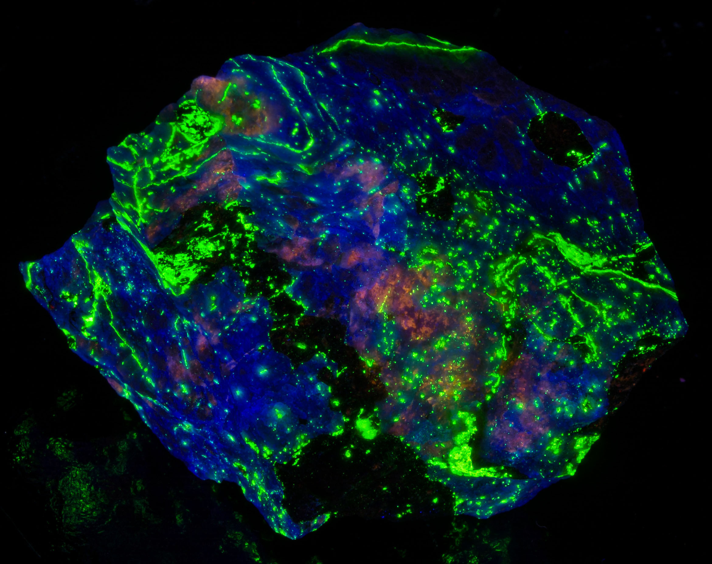
Cataract surgery can produce noticeable changes in the way fluorescent minerals—and colors in general—appear. These changes result primarily from removing the eye’s natural crystalline lens, which typically becomes progressively yellow with age, and replacing it with an artificial intraocular lens (IOL).
The yellowed natural lens acts as a filter, reducing the amount of short-wavelength light reaching the retina. After cataract surgery, more violet and blue light can reach the eye, depending on the type of replacement lens used. Many people therefore notice that whites appear whiter and blues appear brighter or more intense. Fluorescent minerals with blue or violet emission may become substantially easier to see. For example, blue-fluorescing hardystonite, which may have been difficult to see through an aged natural lens, can appear dramatically brighter after lens replacement.
Some people experience temporary cyanopsia, in which the world appears unusually blue or bluish-white following surgery. This generally results from the sudden increase in short-wavelength light reaching the retina, followed by adaptation of the visual system over time. Although the initial blue tint may diminish, improved perception of blue and violet colors can remain.
The type of IOL can make a difference. Modern IOLs generally include UV protection, typically blocking wavelengths below approximately 400 nm to help protect the retina. Some IOLs also include additional blue-light filtering, giving them a slightly yellow appearance and reducing transmission in the violet and blue portion of the visible spectrum. Other lenses provide greater transmission down toward approximately 400–410 nm and may be preferred where accurate color discrimination is important.
For fluorescent mineral collectors, this difference can be significant. A clearer IOL with greater short-wavelength transmission may allow more of the violet or near-visible emission from a mineral to be seen. However, this also means that poorly filtered UV flashlights—particularly lights operating around 395 nm—may appear much brighter and more intrusive after surgery. The visible purple light from such lamps can overwhelm or reduce the apparent contrast of fluorescence.
A properly filtered 365 nm UV light should therefore be especially useful for fluorescent mineral viewing. Good UV filtering reduces the visible violet output from the lamp while allowing the mineral’s fluorescence to stand out. Appropriate UV-blocking eye protection should also be used when viewing minerals under shortwave UV.
Not all replacement lenses have identical spectral transmission. Before surgery, a patient may be offered different IOL types, including standard clear lenses and blue-light-filtering lenses. Anyone for whom color perception is particularly important—including artists, photographers, and fluorescent mineral collectors—may wish to discuss the spectral characteristics of available IOLs with their ophthalmologist before surgery.
It is also worth noting that cataract surgery outcomes and lens choices vary. Not every patient receives the same type of IOL, and additional factors—including whether one or both eyes are being treated—may affect the choice of lens.
In short: cataract surgery often makes the blue and violet end of the spectrum more visible by removing the yellow filtering effect of an aging natural lens. This can make some fluorescent minerals appear brighter and more colorful, but it can also make unwanted visible output from poorly filtered UV lamps much more noticeable. The exact effect depends partly on the spectral transmission characteristics of the replacement IOL.
This article discusses visual observations and optical effects relevant to fluorescent mineral viewing and is not medical advice. Questions about cataract surgery or IOL selection should be discussed with a qualified ophthalmologist.