
Agrellite, Baratovite, and Albite – Dara-i-Pioz, Tajikistan
Contributed by: Michael Crawford
Date: Aug 2nd, 2026
Locality: Dara-i-Pioz Massif, Districts of Republican Subordination, Tajikistan (See on Mindat)
Size: 7 x 13.5 cm
Description:
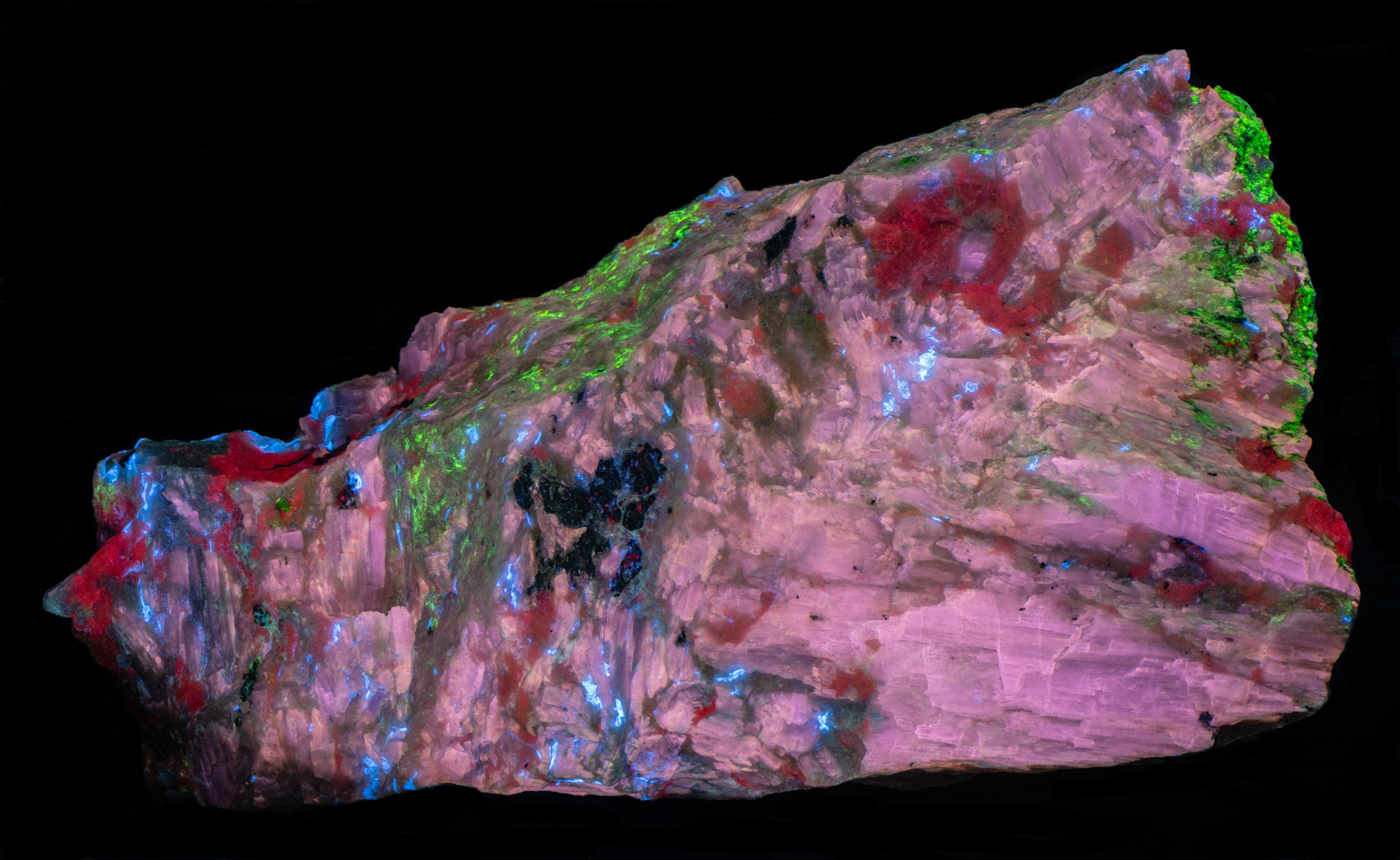
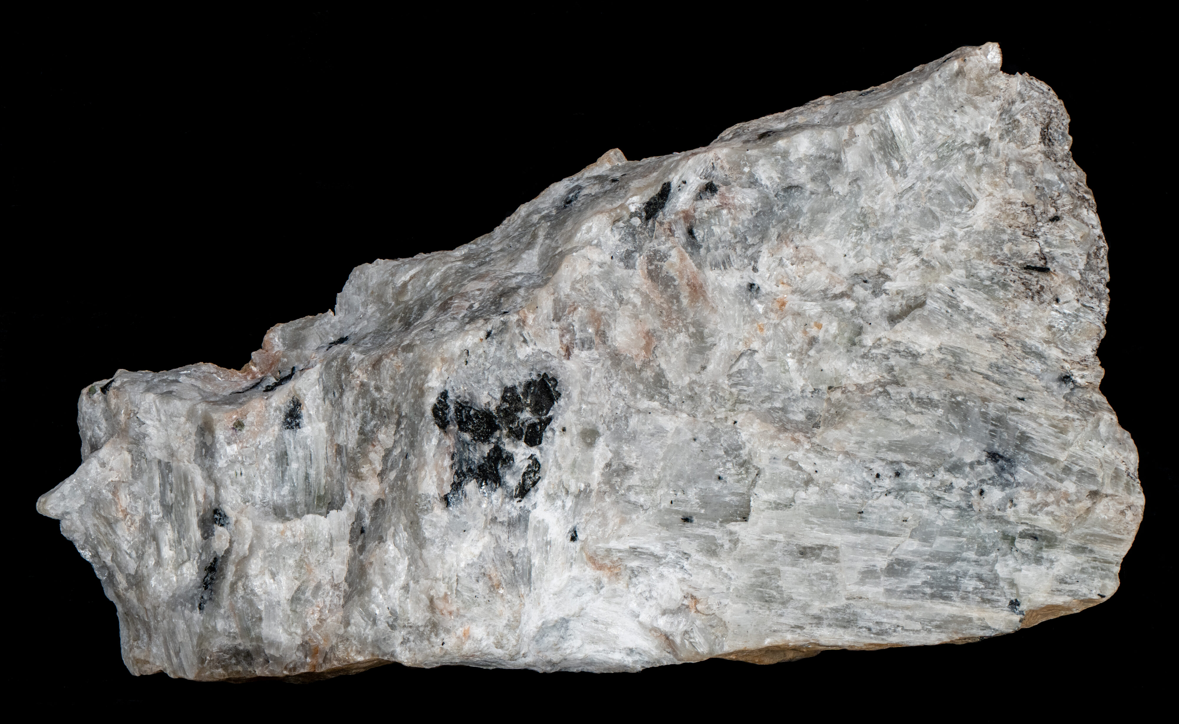
This specimen contains four fluorescent minerals: agrellite (NaCa2Si4O10F), albite (Na(AlSi3O8)), scattered grains of baratovite (KCa7(Ti,Zr)2Li3Si12O36F2) and opal-AN. The specimen comes from the Dara-i-Pioz Massif in Tajikistan, an unusual granitoid intrusion enriched in boron, lithium, and many cesium-bearing minerals. Dara-i-Pioz is the type locality for 47 minerals, including the baratovite present in this specimen.
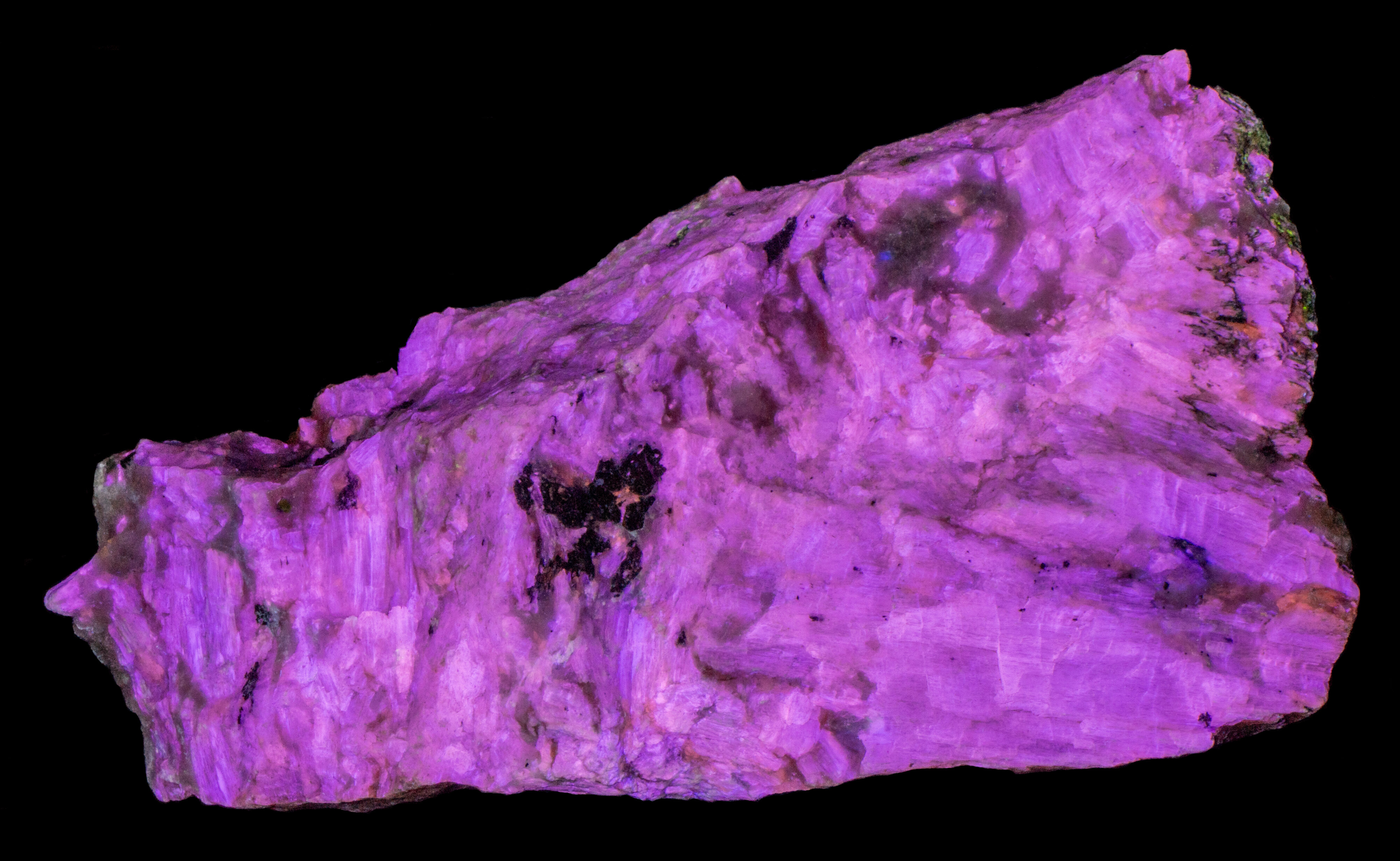
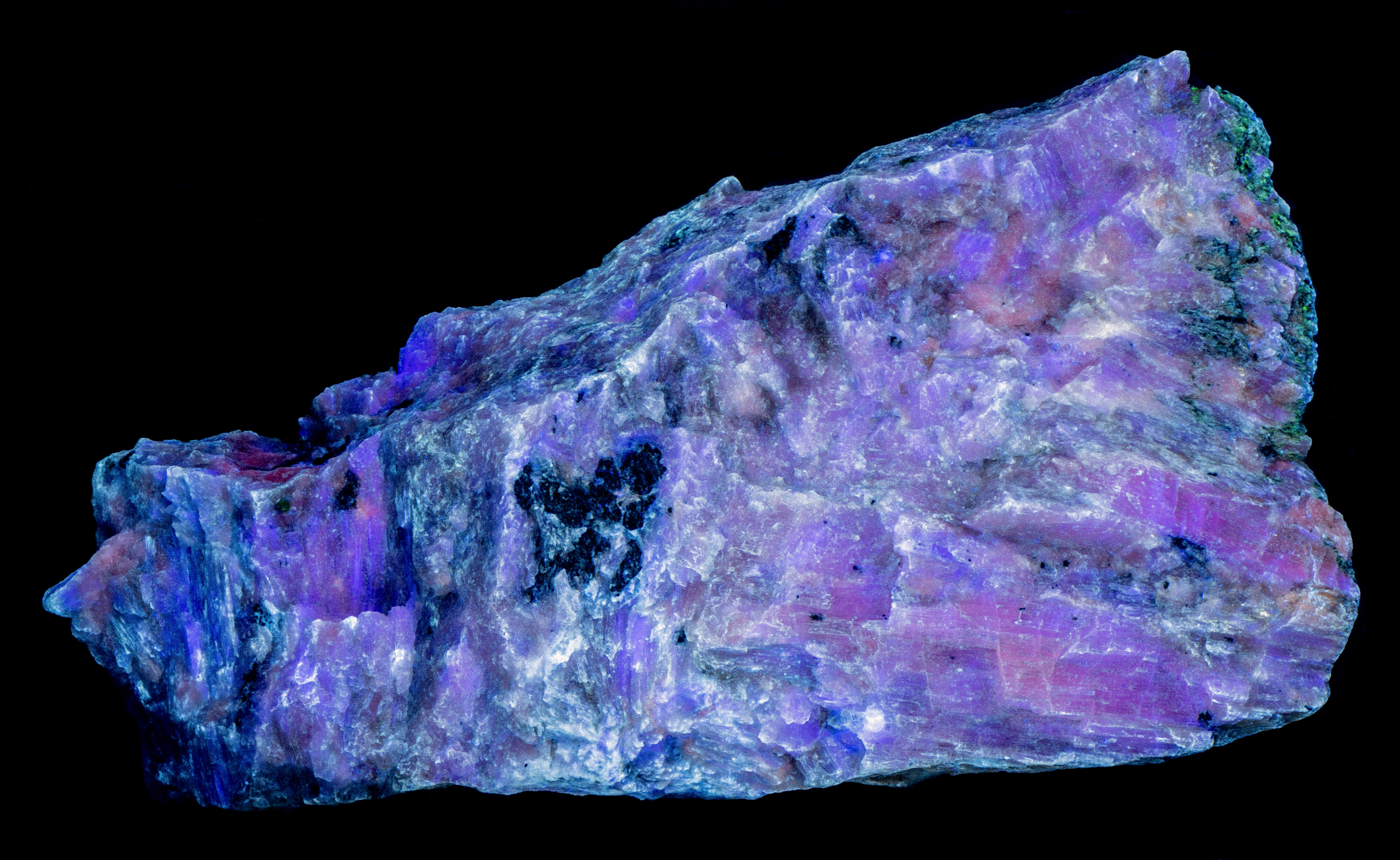
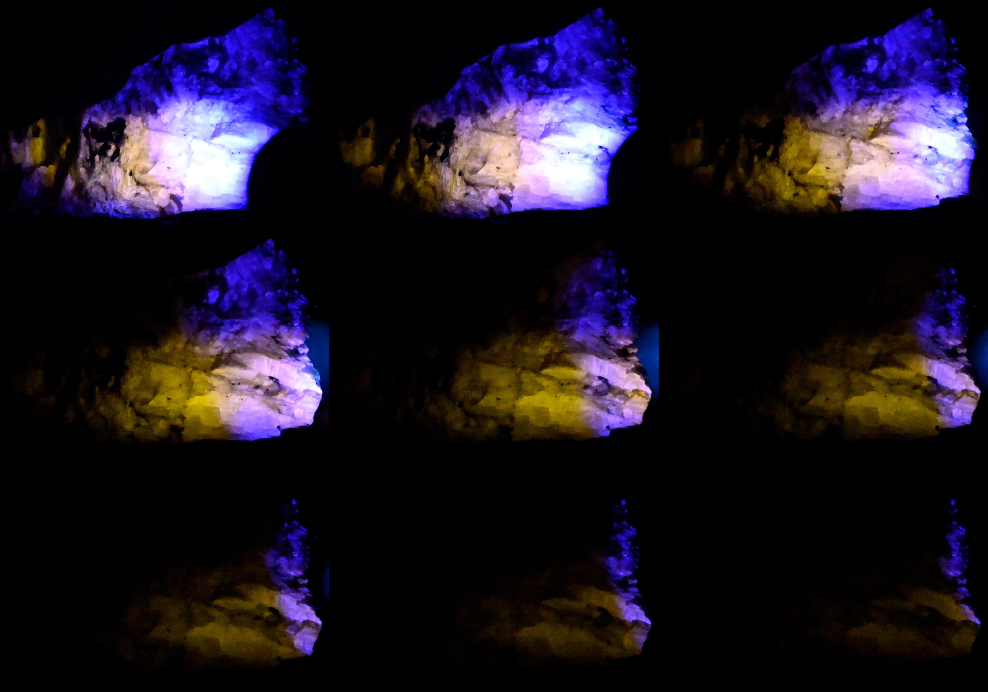
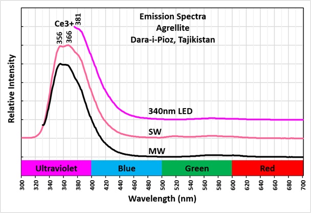
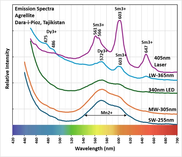
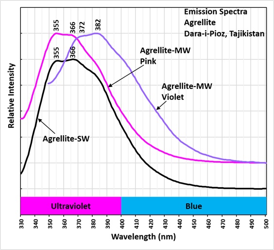
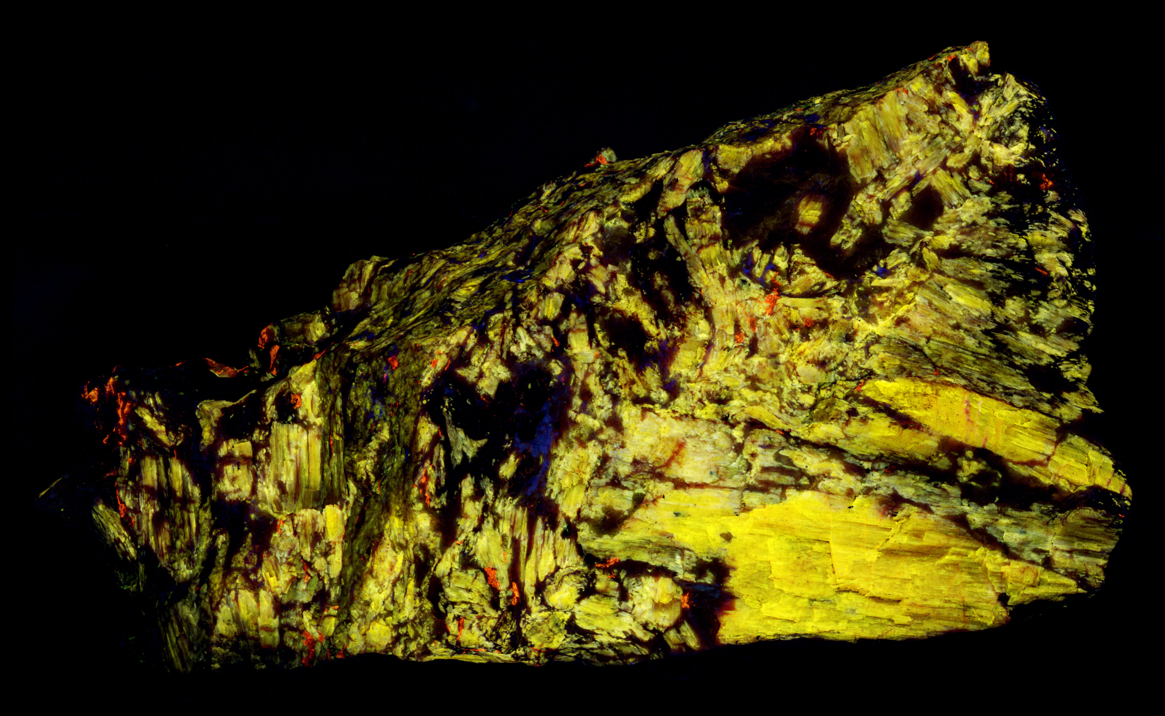
Agrellite appears brighter under a 340 nm LED and shows a deeper pink fluorescence at that wavelength. Its pink response under 340 nm, MW (305 nm LED), and SW (255 nm LED) excitation comes from a combination of very bright ultraviolet fluorescence extending into the visible violet-blue region and weaker yellow-orange visible fluorescence. The SW and MW emission spectra show that the ultraviolet fluorescence is nearly 30 times stronger than the visible yellow-orange emission. This ultraviolet fluorescence is activated by cerium (Ce3+) substituting for calcium in the agrellite structure, producing sharp peaks at 355 nm and 366 nm and an inflection near 381 nm in the emission spectrum.
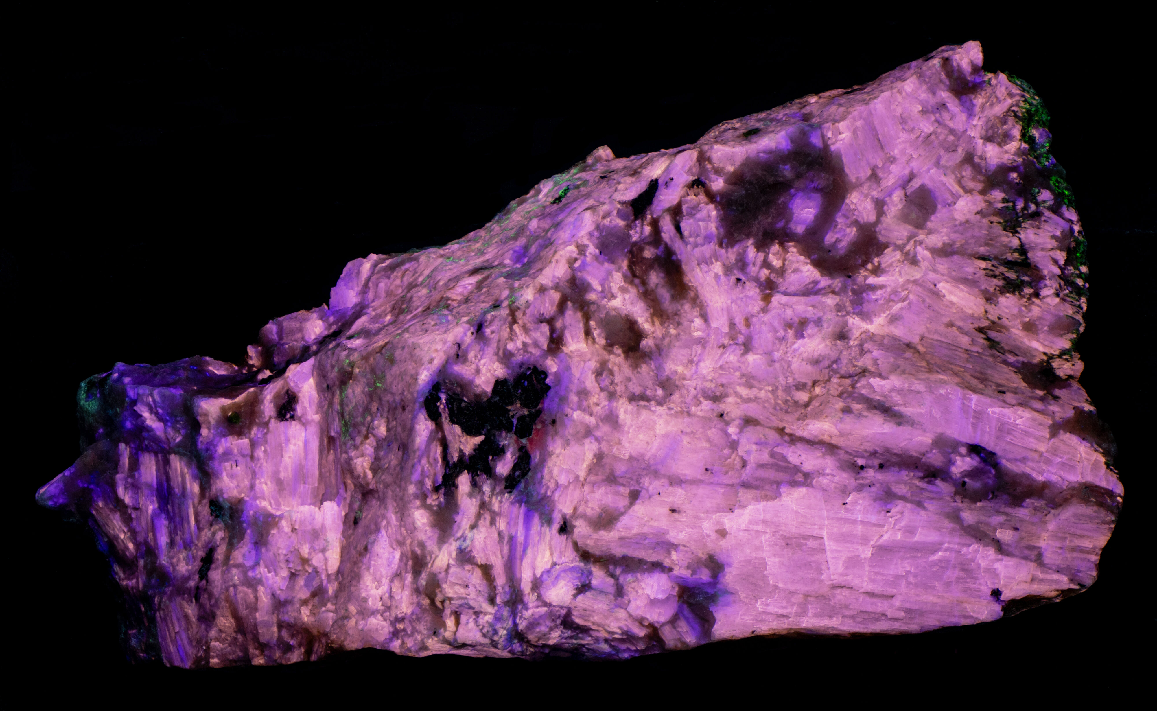
The midwave fluorescence image shows two colors in agrellite: the typical pink and patches of violet fluorescence. In the pink agrellite, the midwave emission spectra show ultraviolet peaks at 355 nm and 366 nm, similar to its shortwave emission. In the violet agrellite, the midwave ultraviolet peaks shift to longer wavelengths, at 372 nm and 382 nm. The cause of this difference is unknown, but possible explanations include variations in cerium concentration, another activator such as europium, changes in atomic coordination, or lattice defects.
Agrellite's visible yellow-orange fluorescence is activated by dysprosium (Dy3+), samarium (Sm3+), and manganese (Mn2+) substituting for calcium. Manganese produces a broad emission centered around 580 nm, with sharper peaks superimposed on the broad peak at 572 nm and 603 nm. The 572 nm peak is activated by dysprosium, while the 603 nm peak is activated by samarium. This double-peak-on-broad-peak pattern appears in the SW, MW, 340 nm, and LW emission spectra. As the excitation wavelength increases, the broad manganese peak becomes less intense. Manganese activation is also indicated by yellow Brief Intense Phosphorescence (BIP), which is strongest when a MW or 340 nm flashlight is swept across the specimen.
Dysprosium and samarium also produce additional sharp peaks in the LW and 405 nm laser emission spectra. Dysprosium adds peaks at 475 nm and 486 nm, while samarium adds peaks at 561 nm, 566 nm, and 647 nm.
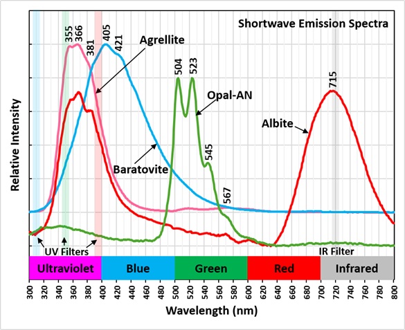
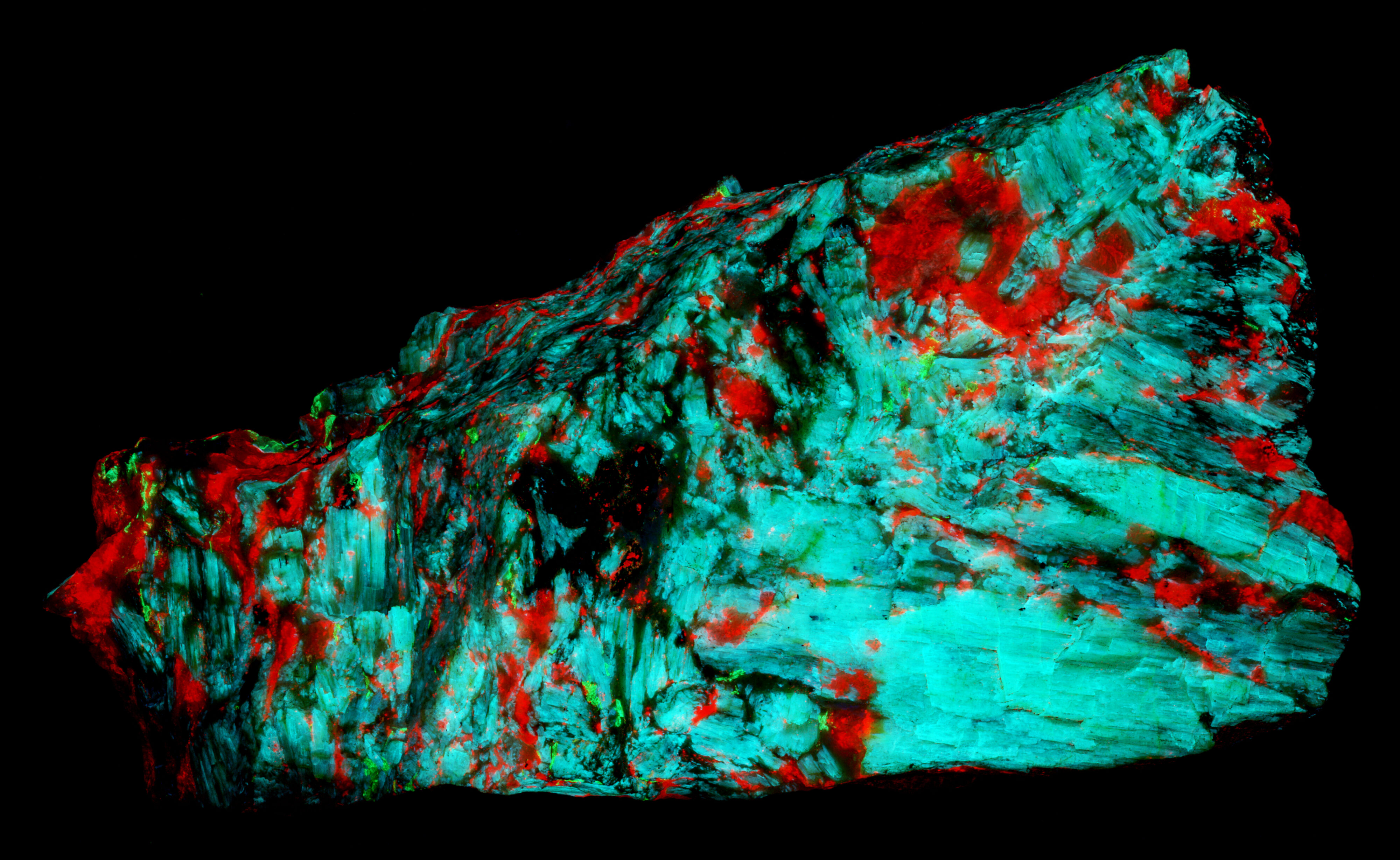
The shortwave fluorescence image reveals three additional fluorescent minerals: light blue baratovite, red albite, and green uranyl-activated opal-AN. Baratovite's shortwave emission spectrum has a double peak at 405 nm and 421 nm, with a shoulder extending into the ultraviolet. Its fluorescence is likely activated by TiO6, which is chemically part of the baratovite structure, making the fluorescence intrinsic. Smaller peaks suggest that a rare earth element, possibly cerium, may also contribute to the light blue fluorescence.
Albite's red fluorescence is activated by ferric iron (Fe3+) substituting for aluminum. Its emission spectrum forms a broad peak centered at 715 nm and also shows ultraviolet fluorescence with an agrellite-like emission signature, indicating that agrellite is mixed with the albite.
The green, fluorescent mineral in the shortwave fluorescent image has an emission spectrum with sharp peaks at 504 nm, 523 nm, and 545 nm, plus an inflection near 567 nm. This pattern is characteristic of uranyl-activated opal-AN.
The false-color image highlights agrellite’s bright ultraviolet fluorescence and scattered baratovite grains. It combines three narrow-band filter images shown on the shortwave emission plot: 310 nm assigned to blue, 350 nm to green, and 394 nm to red. In this ultraviolet false-color image, agrellite appears yellow and baratovite red. A second false-color image combines infrared and ultraviolet images. The image uses a near-infrared band for red, the 394 nm UV bandpass for green, and the 350 nm UV bandpass for blue. In the IR-UV image, albite appears red, baratovite green, and agrellite light blue.
Summary of luminescence responses:
Agrellite (Mindat) (RRUFF)
- Fluorescence under Longwave (365nm LED) UV light: Pink
- Fluorescence under Midwave (305nm LED) UV light: Pink
- Fluorescence under Shortwave (255nm LED) UV light: Pink
- Afterglow after exposure to Midwave (305nm LED) UV light: Yellow
- Fluorescence under Shortwave (255nm LED) UV light: Red
- Fluorescence under Shortwave (255nm LED) UV light: Blue
- Fluorescence under Shortwave (255nm LED) UV light: Green