
Multi-color Fluorescent Specimen from Santa Eulalia, Chihuahua, Mexico
Contributed by: Michael Crawford
Date: Oct 29th, 2025
Locality: Santa Eulalia Mining District, Aquiles Serdán Municipality, Chihuahua, Mexico (See on Mindat)
Size: 10 x 16 cm
Description:
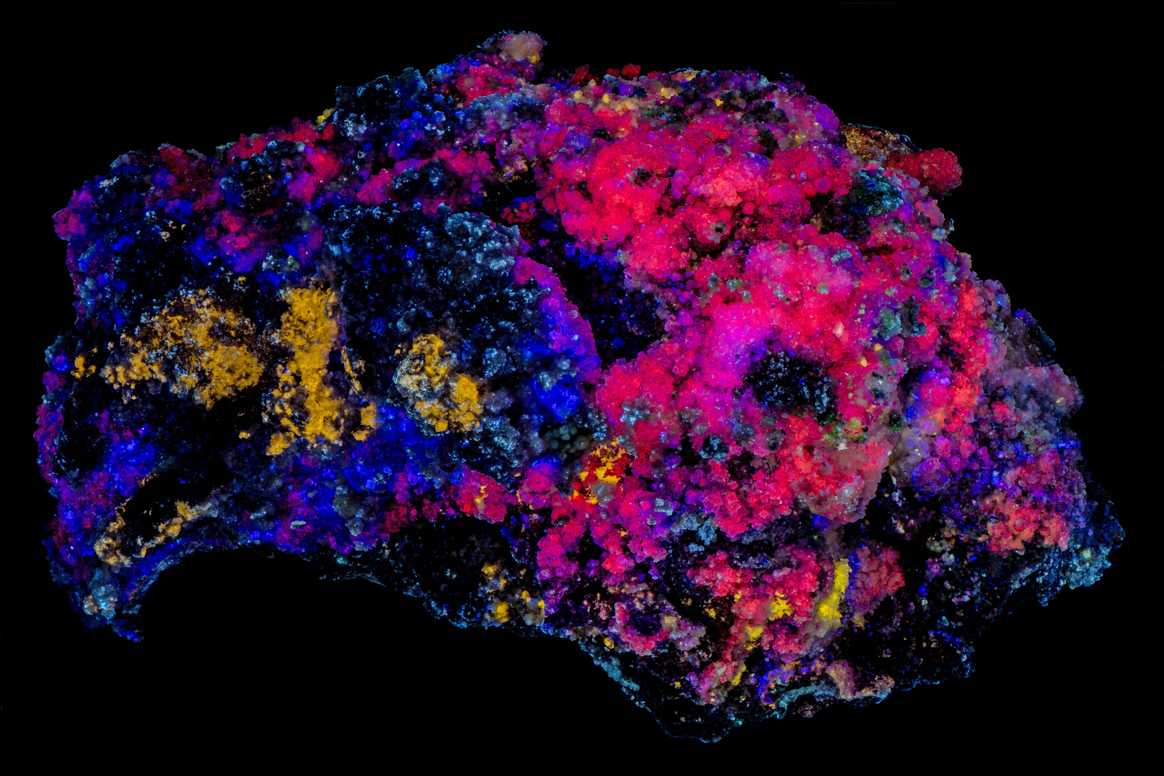
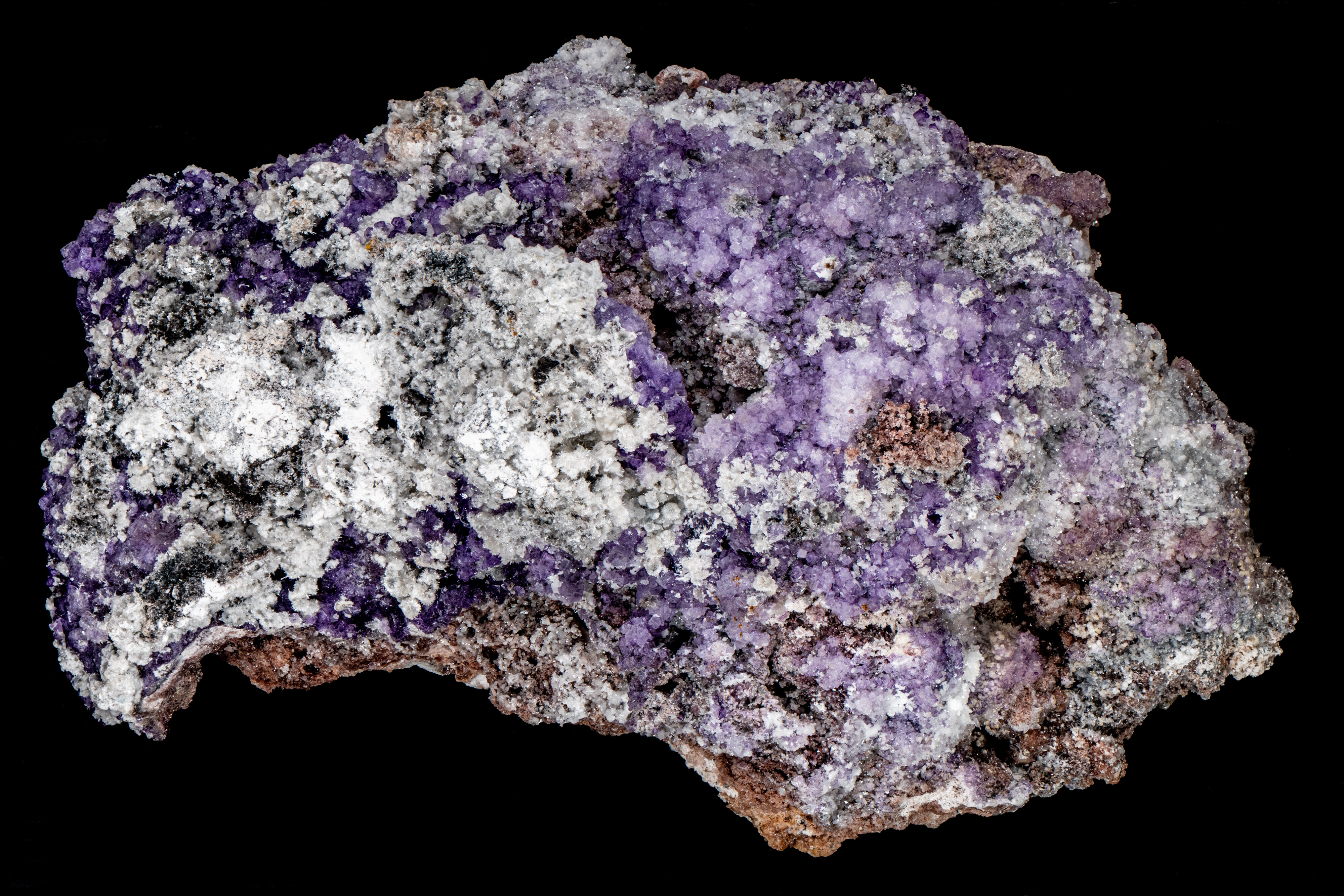
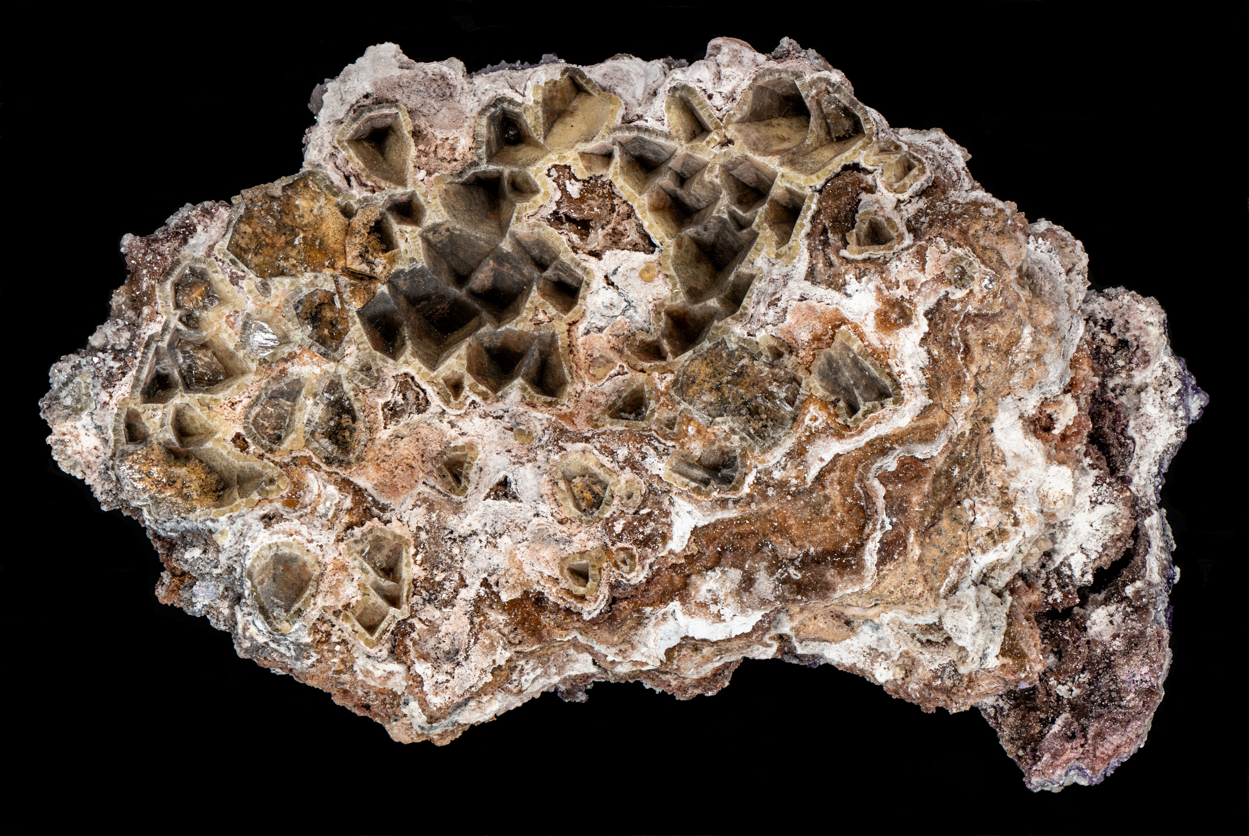
This is a specimen with several fluorescent minerals from the Santa Eulalia District, Chihuahua, Mexico. The specimen contains fluorescent hydrozincite, fluorite, gypsum and calcite.
The fluorite has a purple daylight color and there are five types of fluorescence of the fluorite in this specimen.
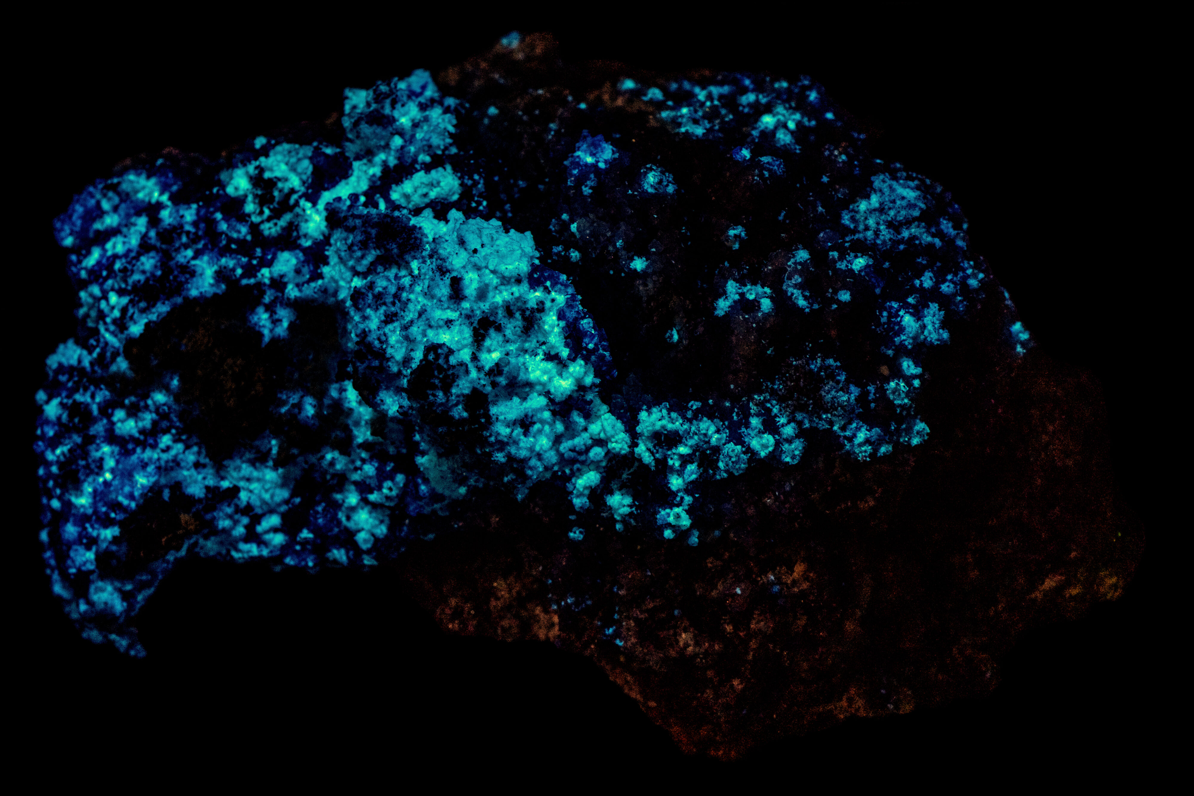
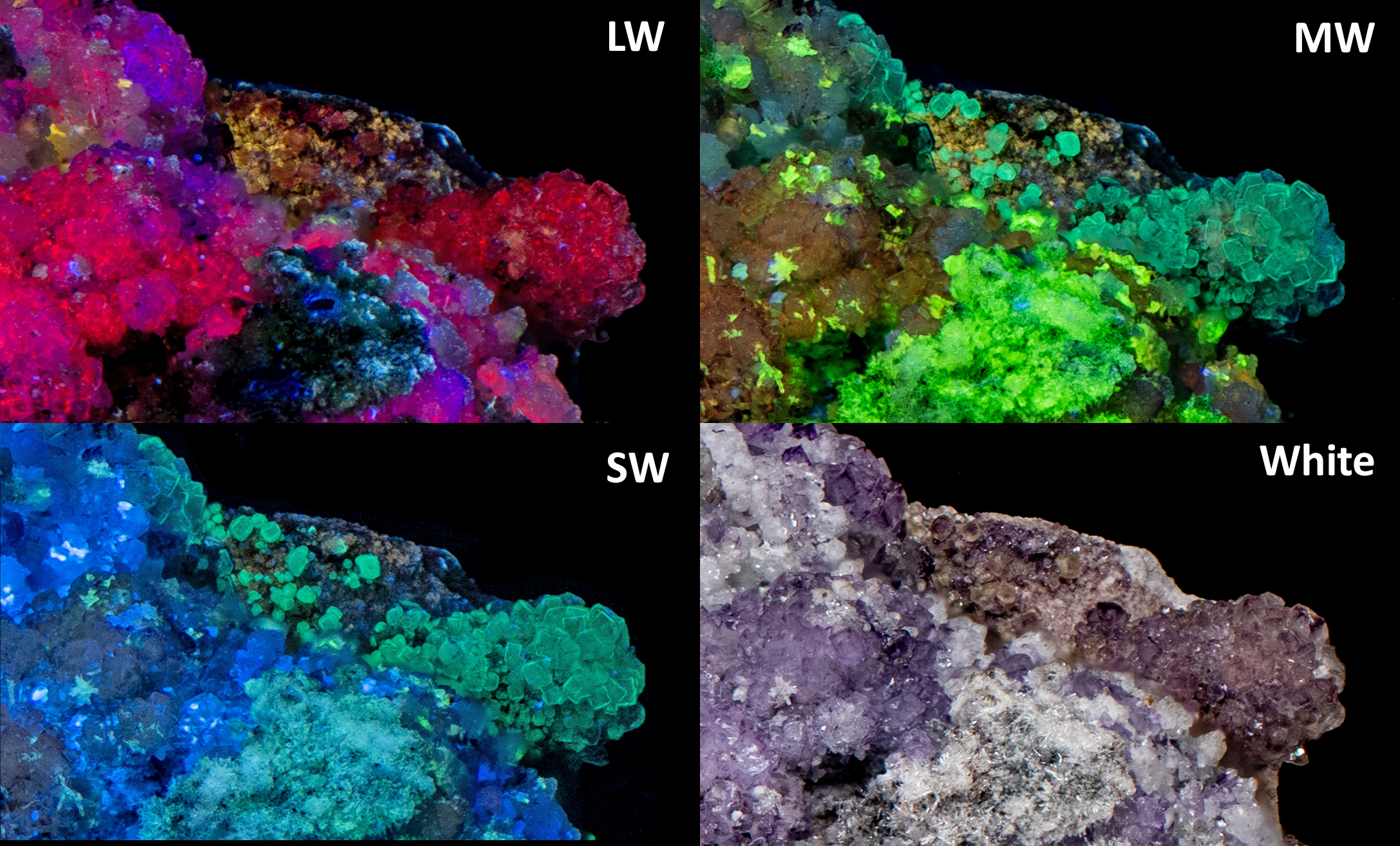
1) Fluorite that fluoresces red and near infrared under longwave and midwave UV light.
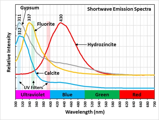
2) Fluorite that fluoresces red and near infrared under longwave and midwave UV light and dim blue under shortwave UV light with strong ultraviolet fluorescence with a peak at 337 nm.
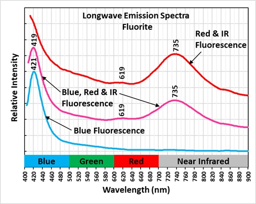
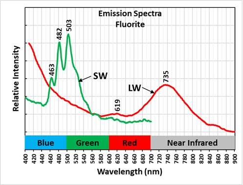
3) Fluorite with blue, red, and near infrared emission peaks at 419 nm, 619 nm and 735 nm
4) Fluorite that only fluoresces blue under longwave UV light.
5) Fluorite that fluoresces red and near infrared under longwave UV light and green under midwave and shortwave UV light
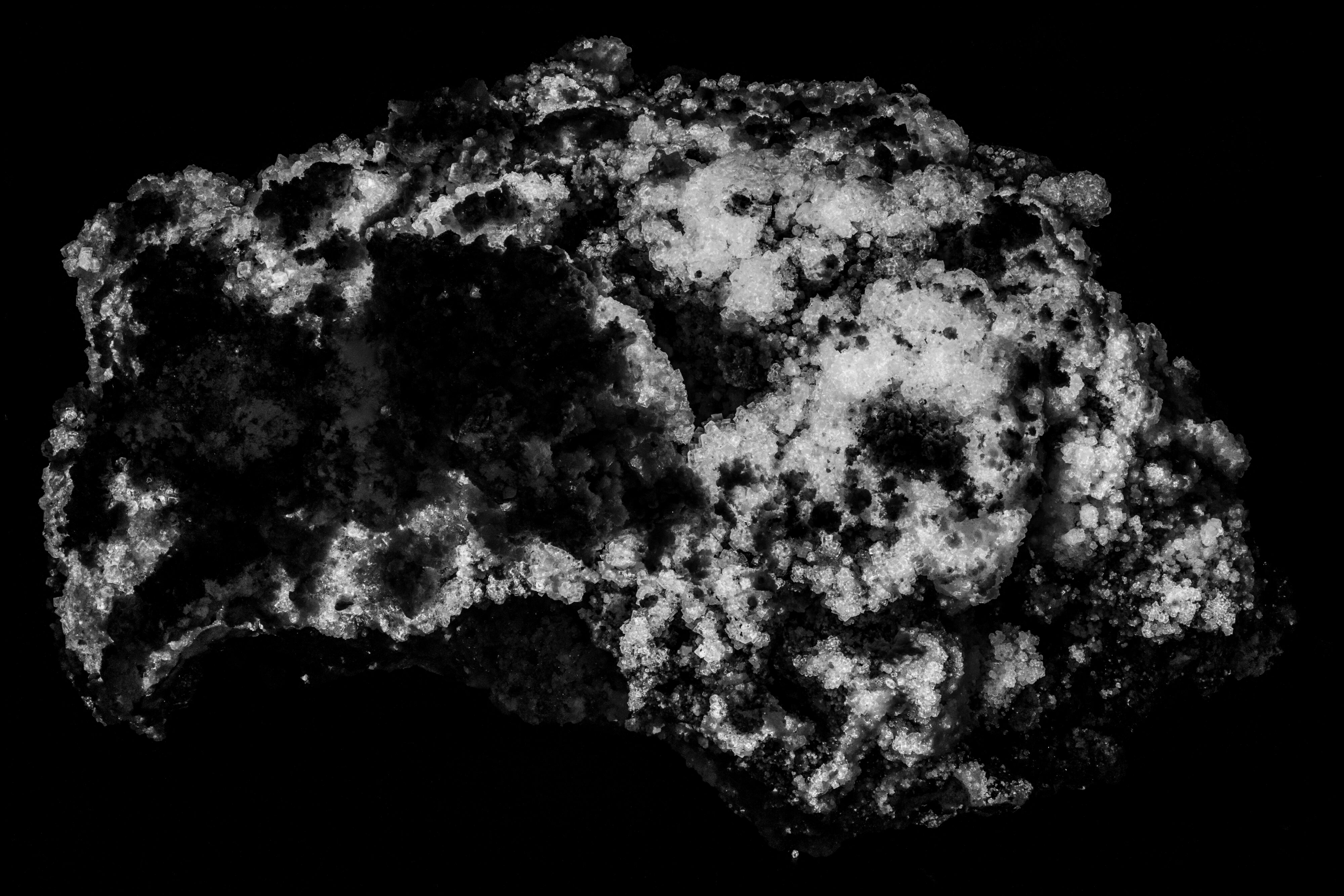
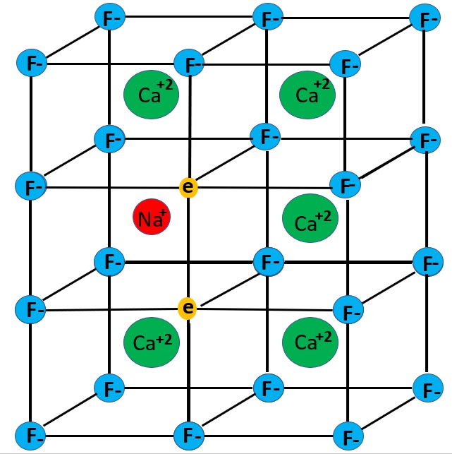
The longwave emission spectrum of the bright red and near infrared fluorescence has a broad peak at 735 nm and a smaller peak at 619 nm. The emission spectra indicates that the red and near infrared fluorescence is a combination of two types of activation. Details of the two mechanisms are discussed in Gaft and others (2020). One mechanism is the presence of M-centers. An M-center forms when two nearby fluorine ions (F-) are missing and are replaced by electrons. The M-center is stabilized by replacement of a calcium ion by a sodium ion. M-centers are formed by naturally occurring radiation that caused the formation of crystal lattice defects. The fluids that deposited the fluorite were radioactive, but generally there is no residual radioactivity. However, some of the fluorite in this specimen, uranyl ions were trapped in the fluorite activating green fluorescence and causing a low level of radioactivity. The radioactivity also caused the purple color of the fluorite. The purple color is caused by colloidal calcium, not the M-center. According to Gaft and others, the M-center activates a broad emission peak with a maximum at 750 nm and a smaller peak at 635 nm.
A second activator of red fluorescence is samarium (Sm2+) replacing calcium. Samarium activation produces a broad peak with a maximum at 730 nm. Emission spectra published in fluomin.org show samarium also activates a peak at 622 nm. The emission peak measured for this specimen peaks at 735 nm. The measured peak is intermediate to the 750 nm peak for the M-center and the 730 nm peak for samarium and suggests the infrared fluorescence is a mix of the two activator mechanisms. The smaller measured peak is at 619 nm. This corresponds to a samarium peak rather the 635 nm M-center peak.
Another property of red fluorescent fluorite is that prolonged exposure to longwave UV light bleaches the red fluorescence. Exposure to shortwave UV light can restore the longwave fluorescence. The number of times that the longwave fluorescence can be restored is limited.
The blue fluorite fluorescence with a longwave emission peak around 420 nm is activated by europium (Eu2+) replacing calcium. The fluorite with the ultraviolet emission peak at 337 nm is likely activated by cerium (Ce3+).
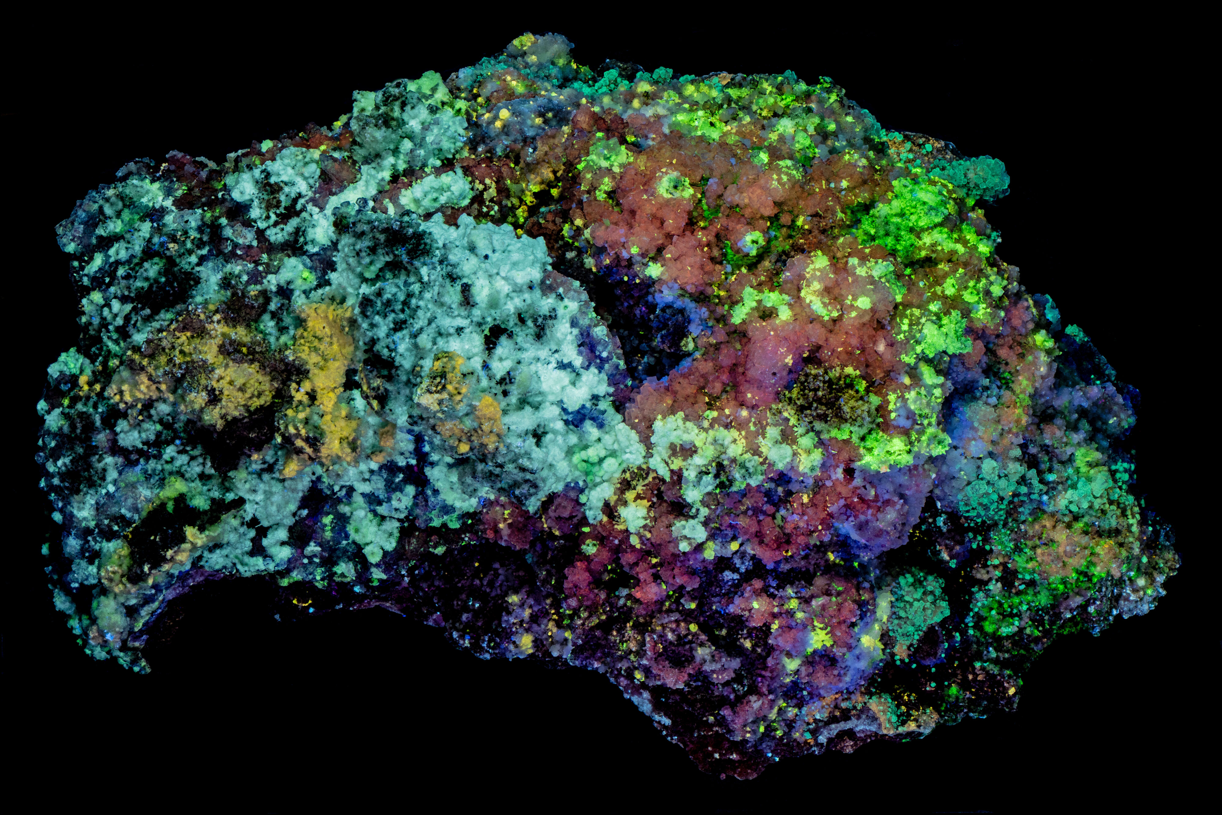
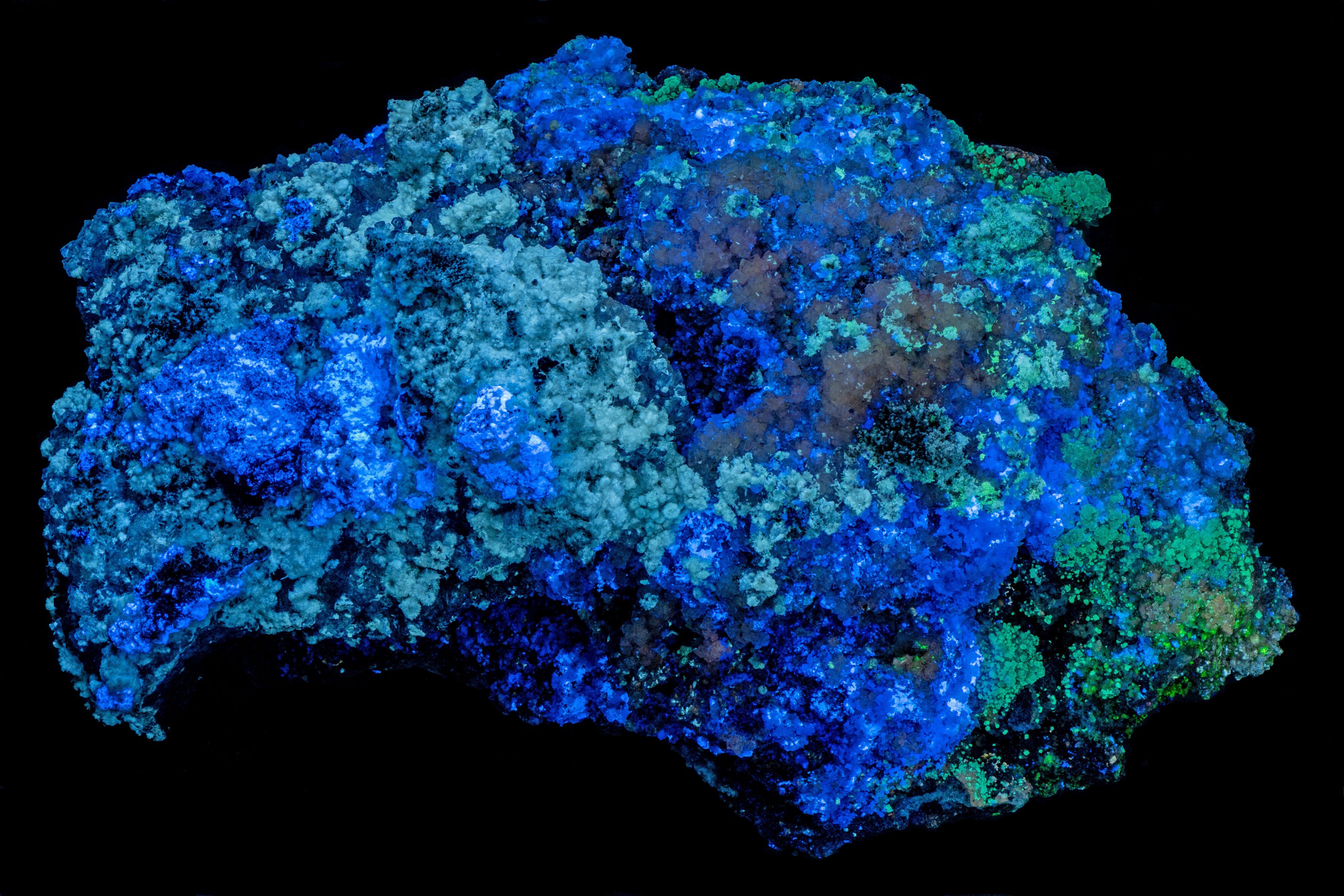
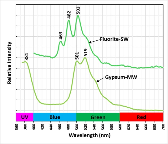
The green fluorite fluorescence is caused by uranyl activation. The shortwave emission spectrum has sharp peaks at 463 nm, 482 nm, and 503 nm. This spectrum is the same as the shortwave emission spectrum of botryoidal fluorite from Fourmile Creek, Fremont County, Colorado. See https://www.mindat.org/photo-1104084.html.
There are three types of gypsum fluorescence in this specimen.
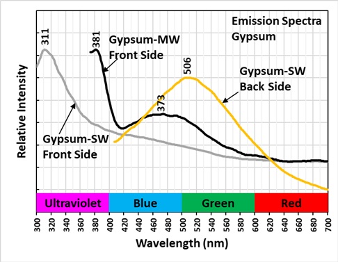
1) Gypsum that fluoresces off-white under midwave UV light, bluish white under shortwave light, and is non-fluorescent under longwave light. It has very long-lasting afterglow from exposure to midwave and shortwave light. This gypsum also has ultraviolet fluorescence with a peak at 311 nm.
2) Gypsum that fluoresces green under midwave UV light, bluish white under shortwave UV light, and is non-fluorescent under longwave light. It also has very long-lasting afterglow from exposure to midwave and shortwave light.
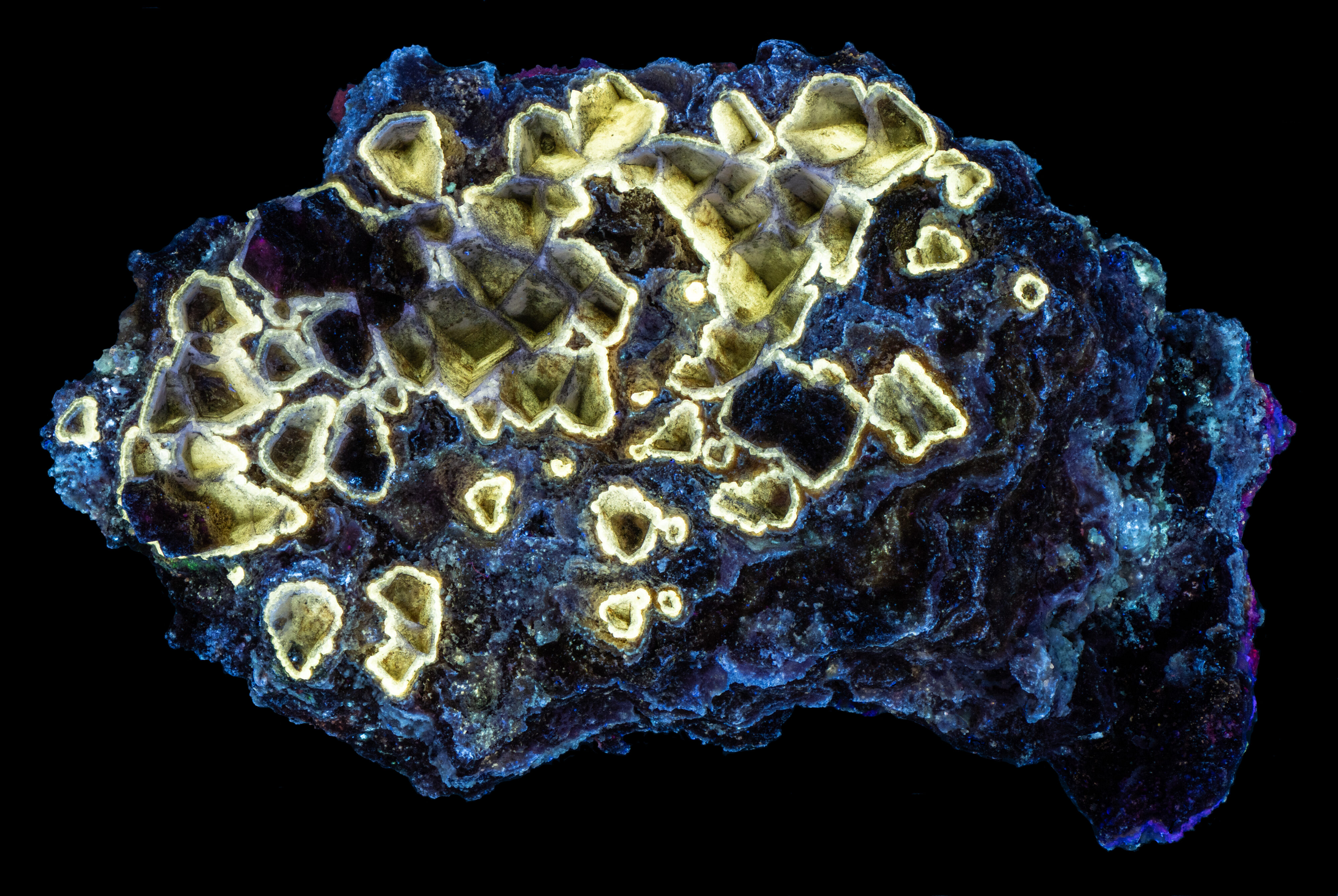
3) The third type of gypsum occurs on the back side of the specimen. It formed a coating on calcite crystals that have dissolved away leaving pyramidal cavities. The fluorescence is yellowish white under all wavelengths of UV light. It is brightest under longwave light. It has faint afterglow.
The green fluorescence in the gypsum is activated by the uranyl ion. The midwave emission spectrum of the uranyl activation has sharp peaks at 501 nm and 519 nm. The off-white and yellow fluorescence as well as the afterglow are likely caused by organic activators. The cause of the ultraviolet peak at 311 nm may also be caused by organics.
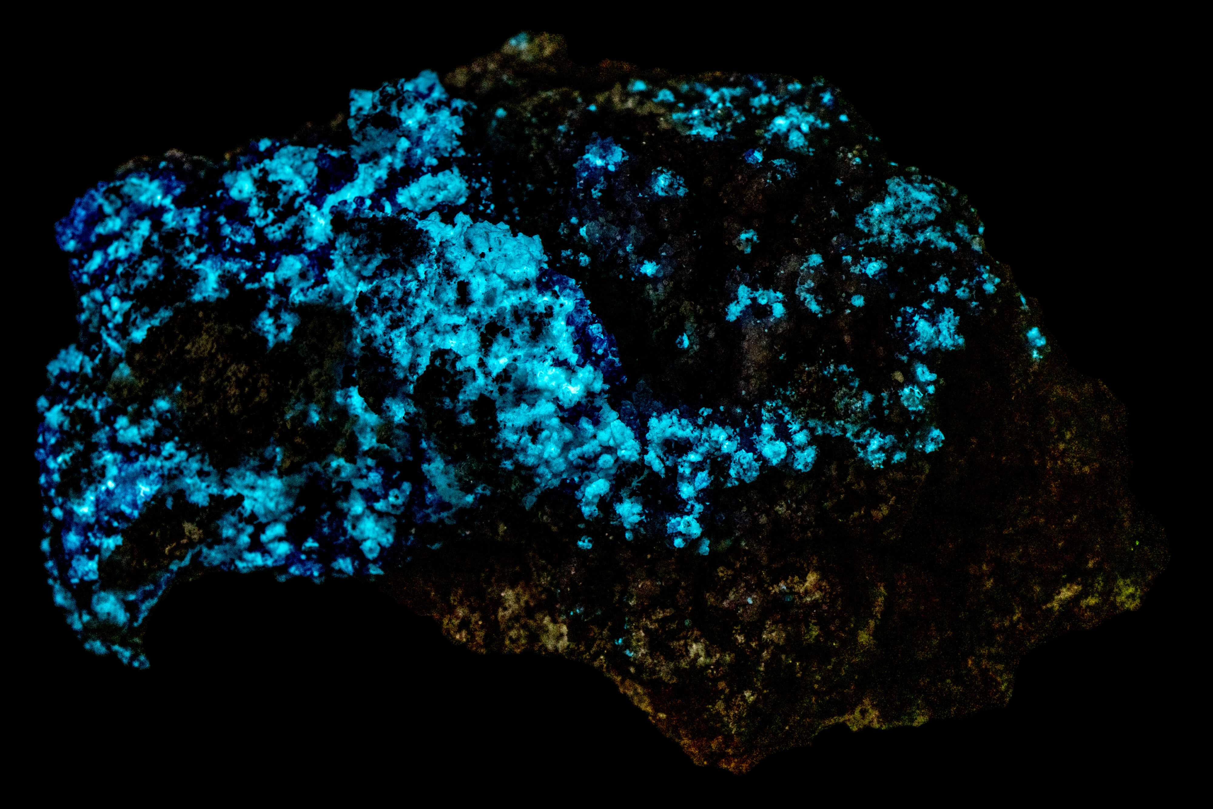
The hydrozincite fluoresces bright yellow under longwave and dimmer yellow under midwave light. The color of the hydrozincite fluorescence changes to bright light blue under shortwave light. The shortwave emission spectrum of blue, fluorescent hydrozincite is a broad peak with a maximum at 430 nm. This fluorescence is activated by lead (Pb2+) replacing zinc. The longwave peak emission peak is at 536 nm for the yellow fluorescence of hydrozincite. The cause of this yellow fluorescence is unknown.
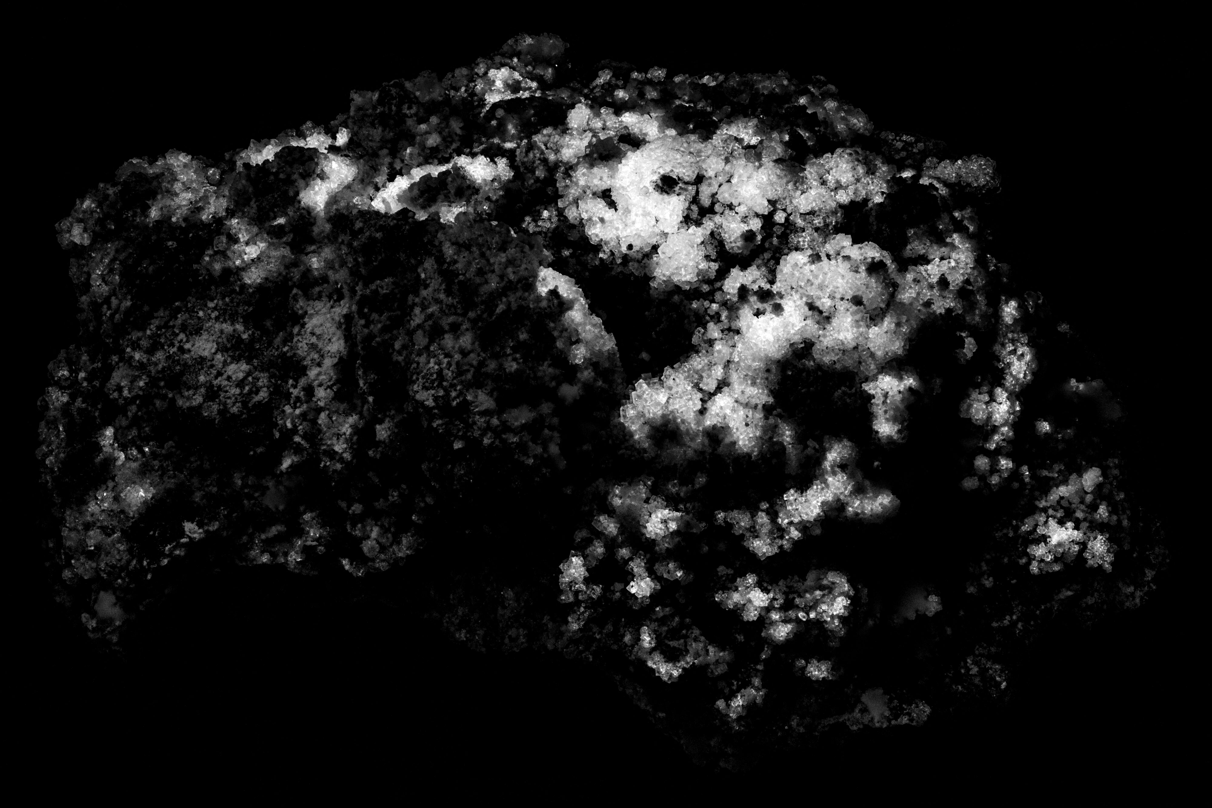
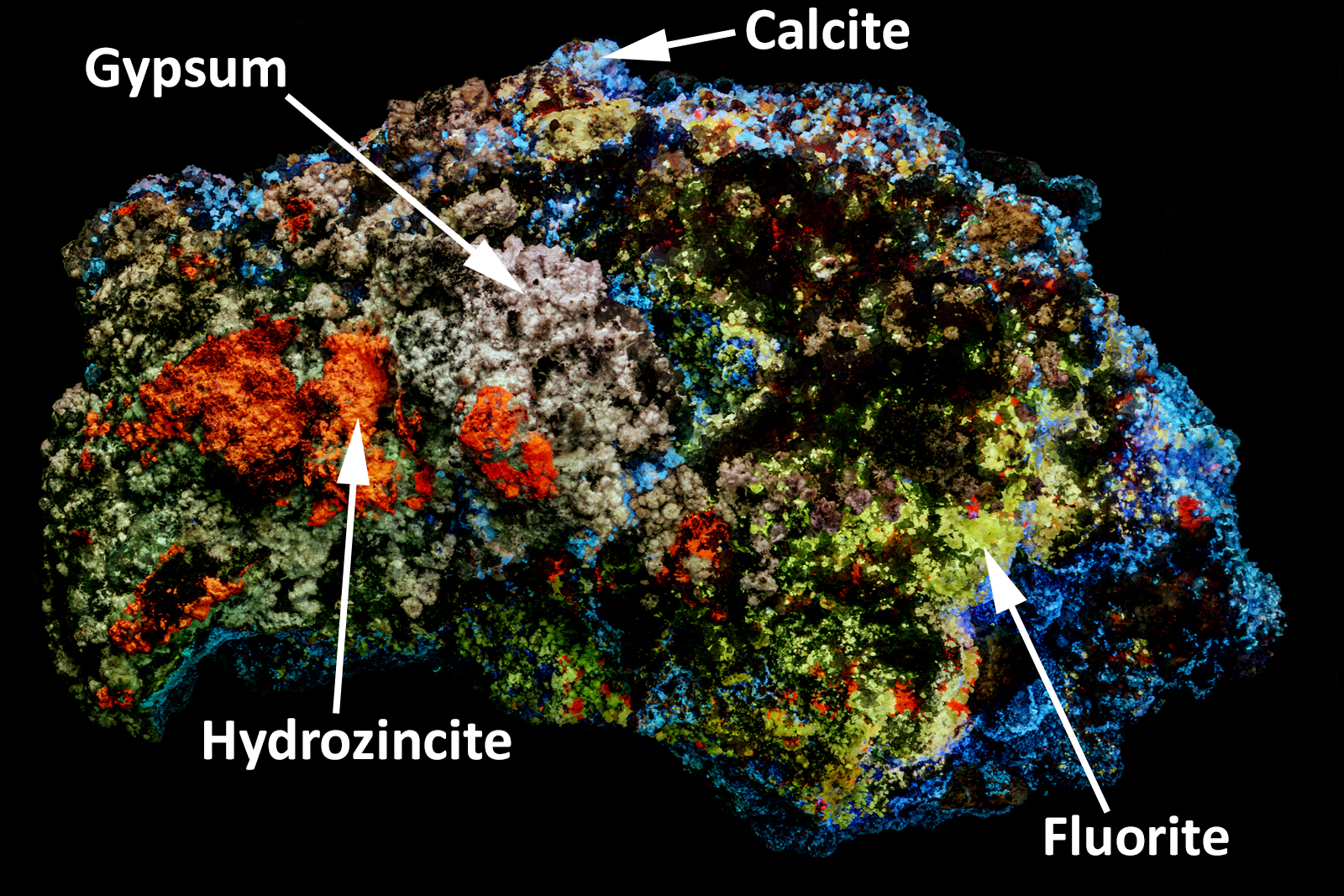
The calcite is most easily recognized in the false color image of ultraviolet fluorescence. The calcite appears light blue in the false color image because of the sharp emission peak at 311 nm. The activation of this fluorescence may be lead (Pb2+) or organics. Further evidence for the light blue areas being calcite is the effervescence produced from a drop of acid.
The false color image is a composite of narrow bandpass images taken at 310 nm, 350 nm and 394 nm. The narrow bandpass filters are shown on the plots of the shortwave emission spectra. The hydrozincite appears red in the false color image. The red is due to the shoulder of blue visible emission that extends into the ultraviolet region. Some of the fluorite appears yellow in the false color image. This fluorite has an emission peak at 337 nm. Gypsum appears off-white in the false color image.
Gaft, Michael & Waychunas, G. & Rossman, George & Nagli, Lev & Panczer, G. & Raichlin, Yosef. (2020). Red photoluminescence and purple color of naturally irradiated fluorite. Physics and Chemistry of Minerals. 47

Summary of luminescence responses:
Fluorite (Mindat) (RRUFF)
- Fluorescence under Longwave (365nm LED) UV light: Red
- Fluorescence under Longwave (365nm LED) UV light: Blue
- Fluorescence under Midwave (305nm LED) UV light: Green
- Fluorescence under Shortwave (255nm LED) UV light: Green