
Terlingua-Type Calcite – Lampasas, Texas
Contributed by: Michael Crawford
Date: Sep 14th, 2026
Locality: Lampasas area, Lampasas County, Texas, USA (See on Mindat)
Size: 8 x 15 cm
Description:
This is a Terlingua-type calcite specimen from the Lampasas area, Texas. Lampasas is 350 miles east-northeast of Terlingua, Texas. The geologic setting of Lampasas is different compared to Terlingua. Terlingua type calcite likely forms from low temperature hydrothermal fluids. The longwave pink fluorescence disappears when specimens are heated between 230oC and 500oC (Waychunas, 2012, Gaft and others, 2008; McCann, 1995). The formation temperature is likely less than 230oC. There are Tertiary age intrusive rocks at Terlingua that could provide a heat source for hydrothermal fluids that deposited the calcite in vertical solution cavities of Cretaceous age limestones (Yates and Thompson,1959). At Lampasas, the calcite is also from Cretaceous age limestones, but there are no nearby intrusive rocks, so the source for heated hydrothermal fluids is unknown.
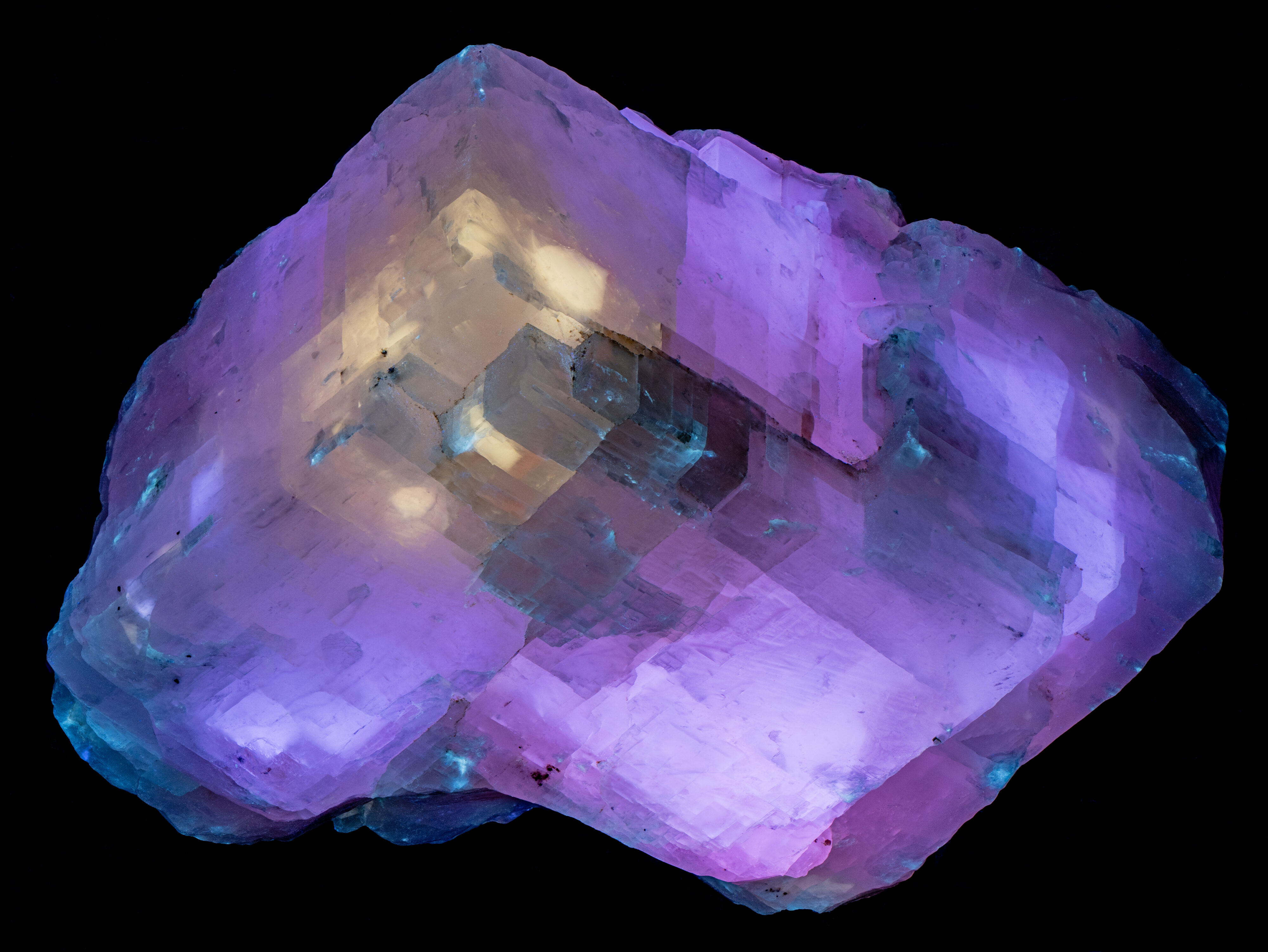
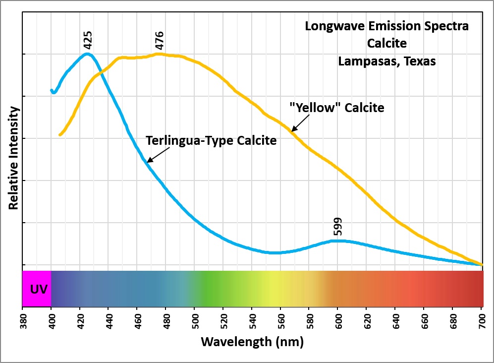
Terlingua-type calcite is characterized by bright blue-white fluorescence under shortwave UV, a bright, persistent blue afterglow, and bright pinkish-orange fluorescence under longwave UV. Its longwave emission spectrum has peaks at 425 nm in the violet-blue region and 599 nm in the orange region; together, these emissions produce the pink color. The violet and orange fluorescence is attributed to radiation-induced lattice defects, likely associated with different rare-earth elements that generate the two peaks (Waychunas, 2012; Gaft and others, 2008). The specific rare earth elements remain unidentified. Radiation causing the lattice defects probably came from uranyl ions in the hydrothermal fluid. Limestone typically has very low concentrations of uranium or other radioactive elements. At Terlingua, the intrusive rocks could be a source for uranium and radioactive hydrothermal fluids. At Lampasas, the source for radioactive fluids is not easily explained.
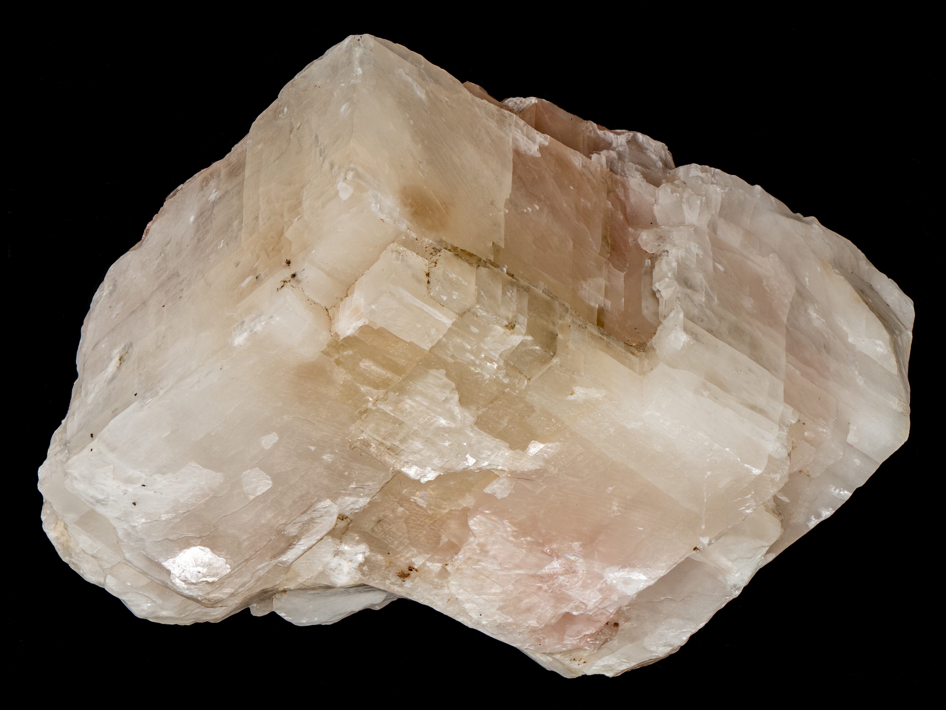
This specimen is composed of pink and white rhombs bounded by cleavage planes. Three directions of rhombohedral cleavage planes are clearly visible in the calcite. The rhombohedral cleavage results from alternating layers of calcium ions and planar carbonate groups (CO3) tilted relative to the crystallographic C-axis. Some of these cleavage faces define rhombohedral zones of different fluorescent colors and intensity. Robbins (1994, 1996) described this zoning in Challenger Cave calcite. He proposed a hypothesis that an activator impurity may have been initially concentrated in some portion of the calcite rhomb and may have spread by diffusion until it was stopped by cleavage planes that were microscopically open, thus barring the way to the additional spread of the activator (Robbins, 1996). Robbin's hypothesis preceded the identification of lattice defects coupled with rare-earth elements as the activators of Terlingua-type fluorescence (Gaft and others, 2008; Waychunas, 2012). The microscopic openings in the cleavage planes may instead have halted the propagation of lattice defects, creating fluorescent boundaries.
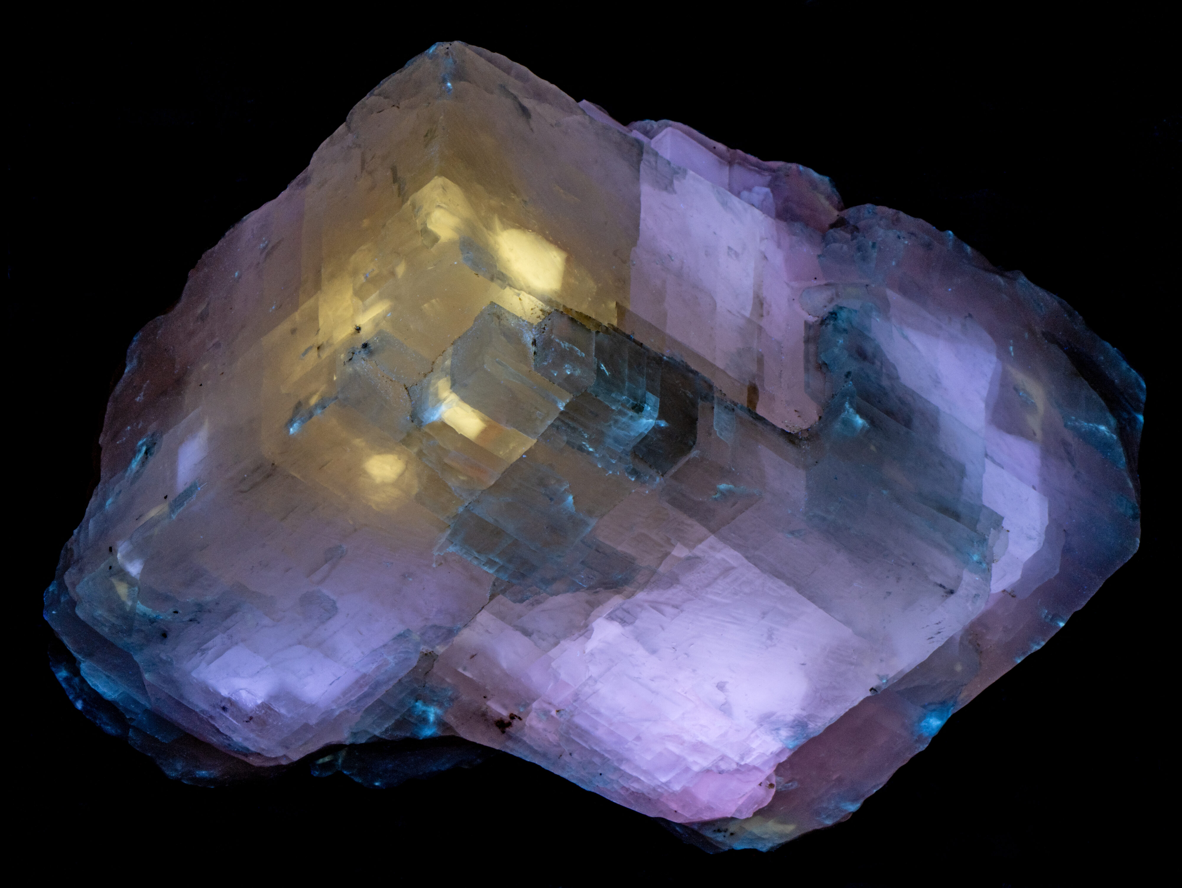
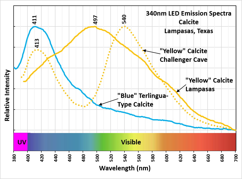
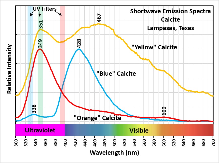
The fluorescent colors in this specimen are paler compared to the standard Terlingua calcite colors. Under longwave, this specimen has a pale pink color with rhombohedral zones of different pink intensities. Standard Terlingua calcite has a more saturated pinkish orange color. Pale-yellow zones are also present and are brightest under longwave excitation. Their emission spectra across all excitation wavelengths suggests organic activators. The 340nm spectrum is different from yellow 340nm emission spectrum of Challenger Cave calcite (another Terlingua-type calcite). The Lampasas spectrum is a very broad peak with a maximum at 497 nm, whereas the Challenger Cave spectrum has two peaks, one at 418 nm and the other at 540 nm. The double peaks suggest lattice defects coupled with rare earth activators.
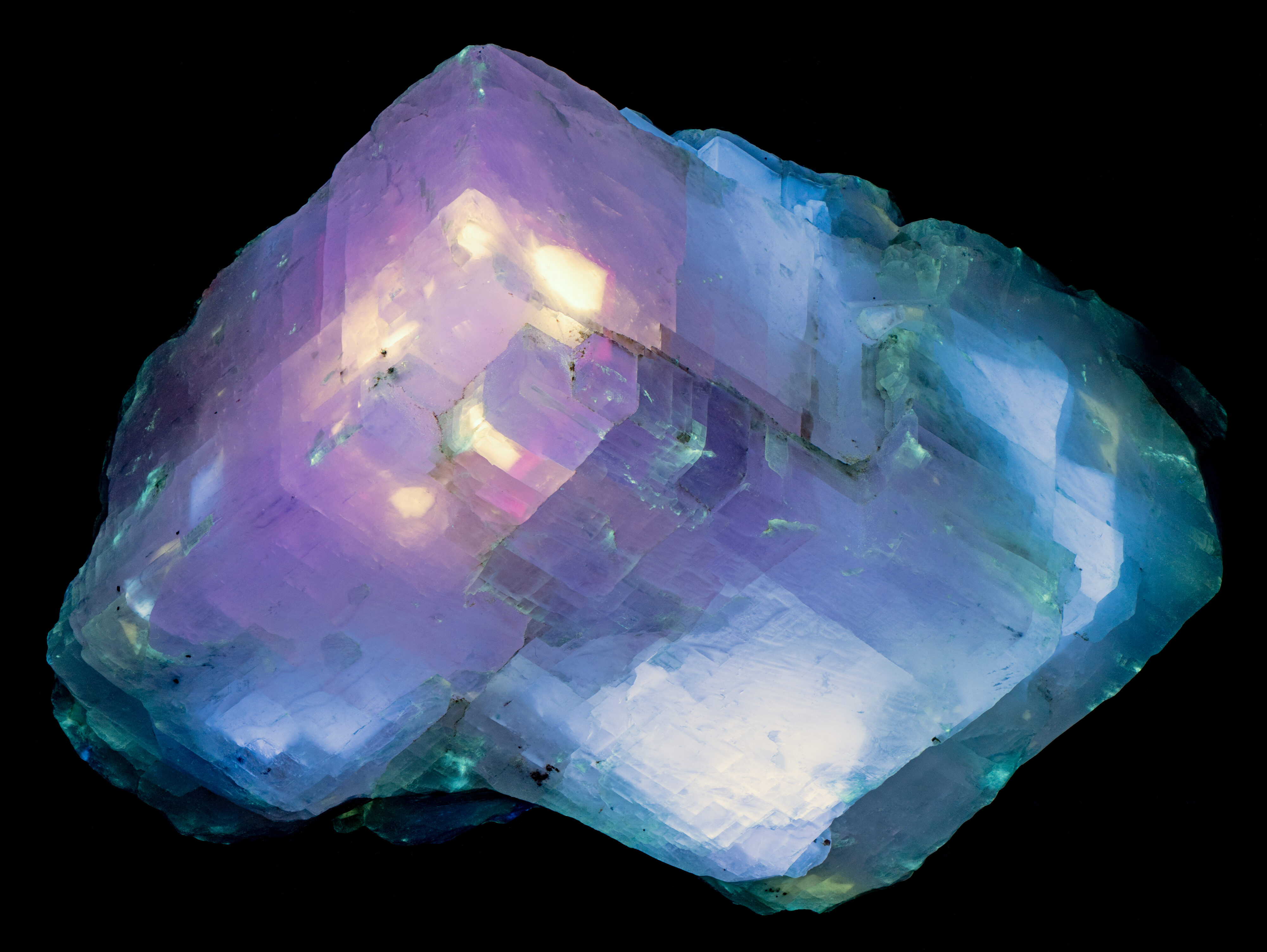
Midwave illumination produces the most colorful fluorescent response, revealing zones of bright blue-white, pale-yellow, and violet-pink fluorescence. The blue-white zones also show a blue afterglow.
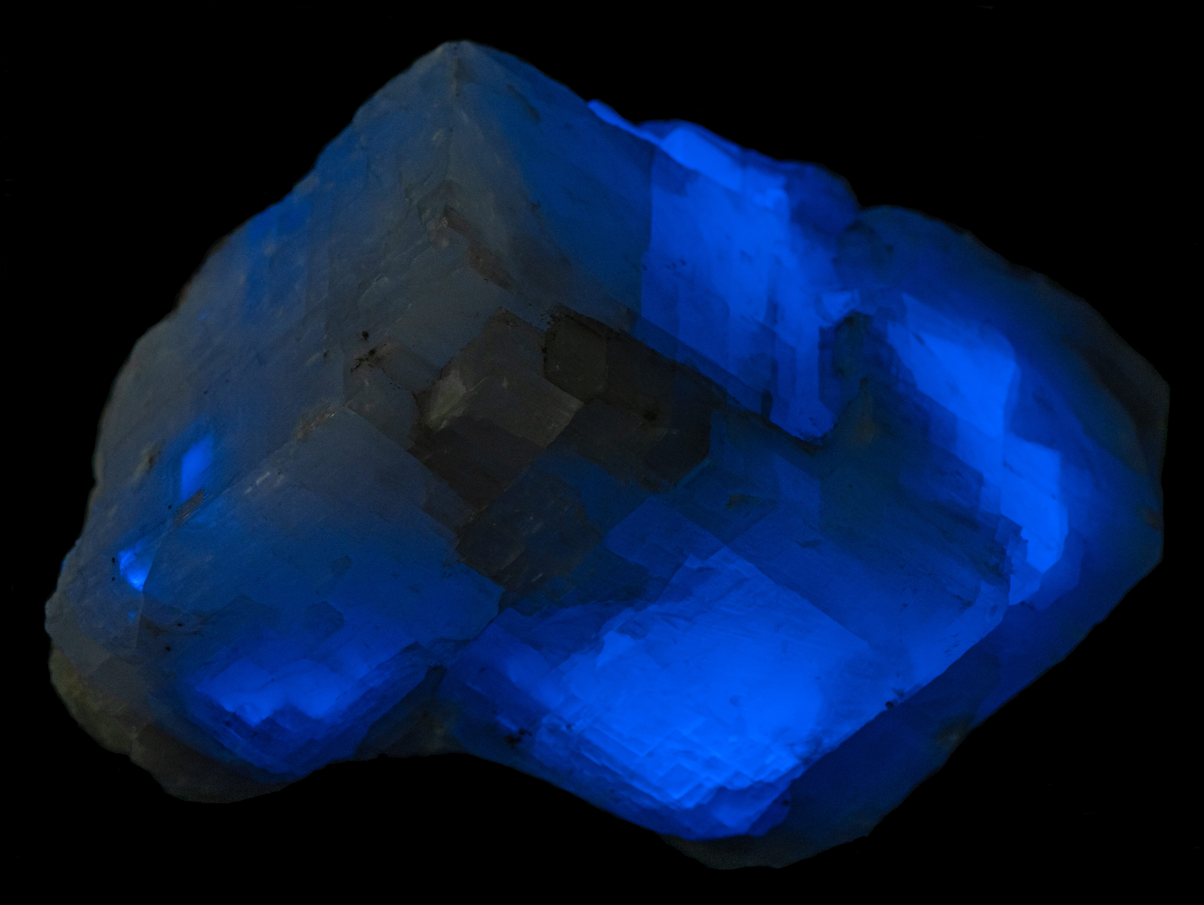
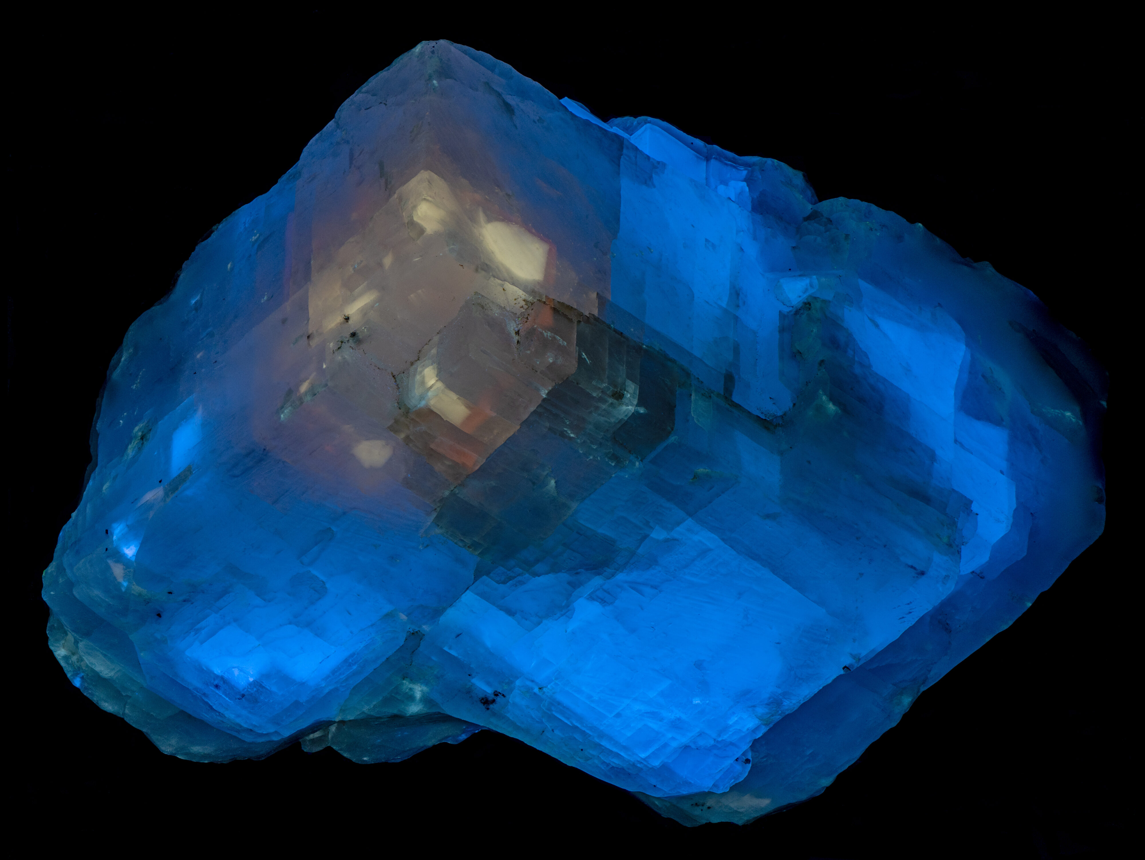
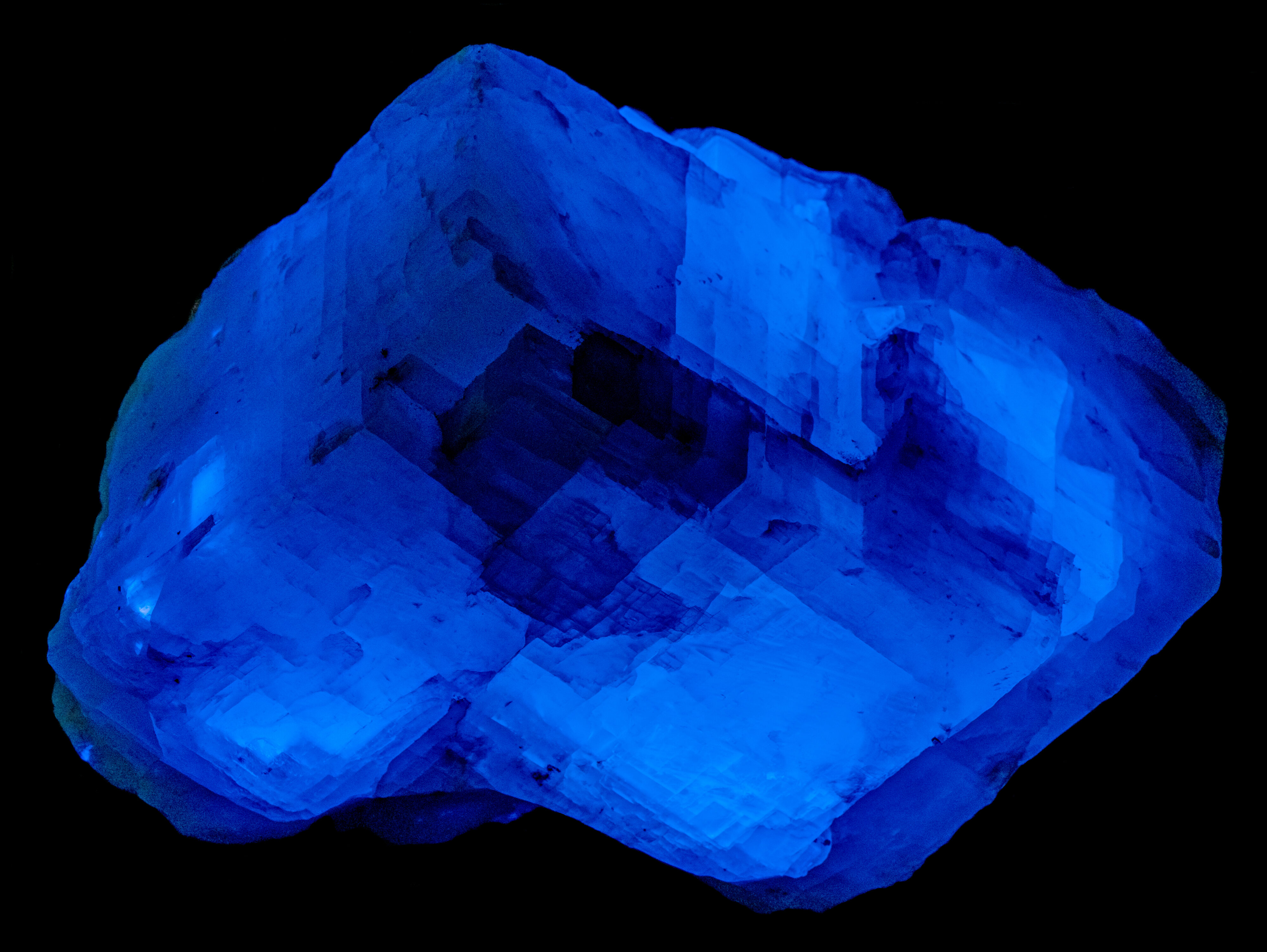
Shortwave illumination from a 255 nm LED also produces a colorful image, with light-blue, pale-yellow, and pale-orange zones. The light-blue zones show a brighter, longer-lasting blue afterglow compared to the midwave afterglow. This afterglow indicates electron traps just below the conduction band. Shortwave UV excites electrons into these traps, and ambient thermoluminescence produces the afterglow (Waychunas, 2012). While the lamp is on, the specimen emits both light-blue fluorescence and blue thermoluminescence. When the light is switched off, the fluorescence stops, but the thermoluminescence continues to slowly release electrons into the conduction band, where they lose some energy and drop to the excited state. As each electron returns from the excited state to the valence band, that energy loss is emitted as a photon of blue light. Temperature affects how quickly electrons leave the traps and, therefore, how long the afterglow lasts. Cooler temperatures slow this release and extend the afterglow.
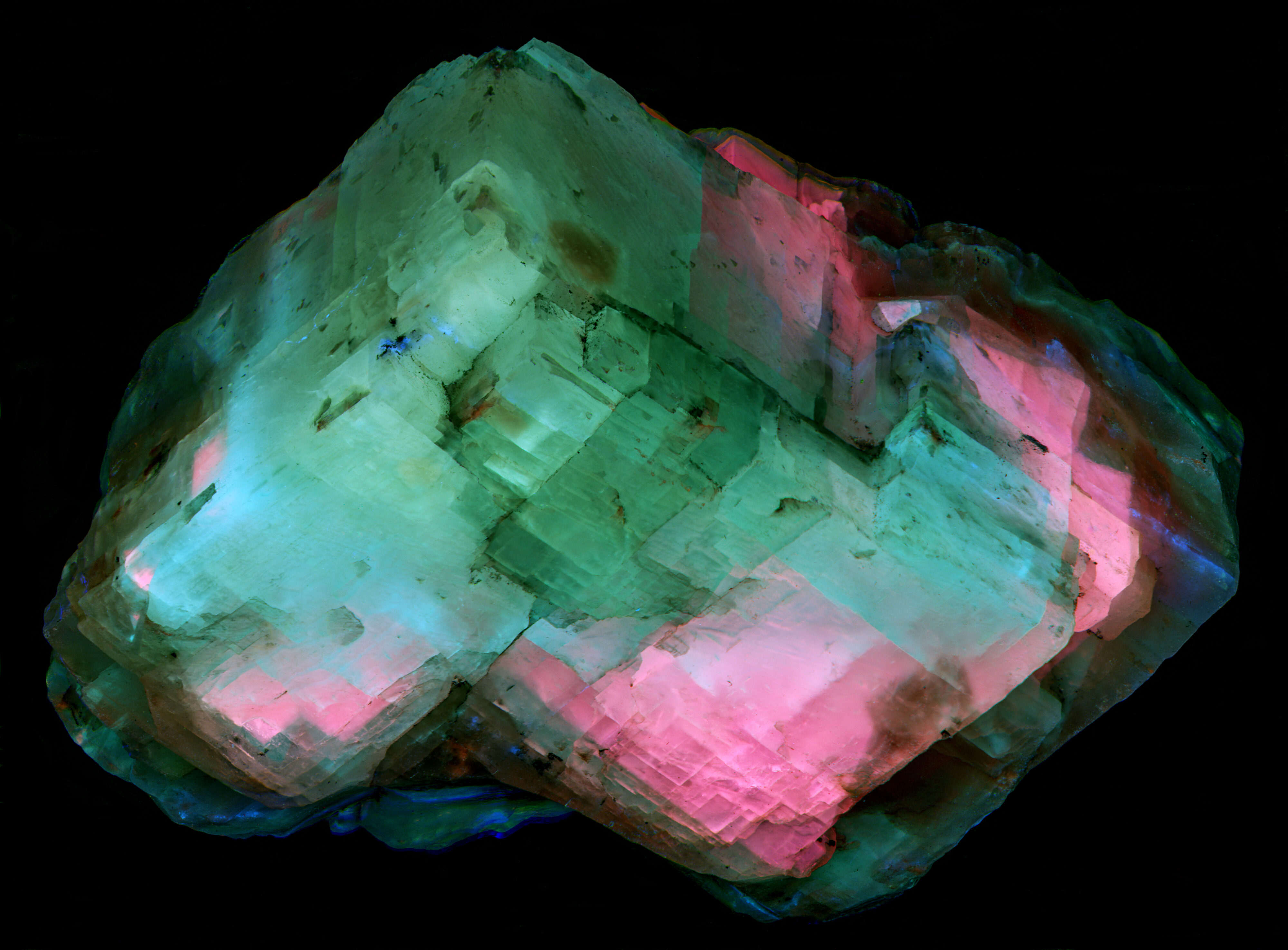
This specimen's shortwave emission spectra contain ultraviolet peaks, an unusual feature in Terlingua-type calcite. The "blue" calcite has a small peak at 338 nm, while the "orange" calcite has a 349 nm ultraviolet peak that is much stronger than its 600 nm orange peak in the visible region. A false-color image of the ultraviolet fluorescence combines narrowband (10 nm bandwidth) images at 394 nm, 350 nm, and 330 nm to red, green, and blue respectively. Green areas in the false color image correspond to the strong 350 nm emission. Pink areas combine red fluorescence from the ultraviolet shoulder of the 428 nm blue peak with blue fluorescence from the small 338 nm peak. Light-blue areas reflect contributions from the 338 and 349 nm peaks. Cerium and europium may activate these ultraviolet emissions.
References:
Gaft, M., Nagli, L., Panczer, G., Waychunas,G., Porat,N., 2008, The nature of unusual luminescence in natural calcite CaCO3, American Mineralogist, V.93, pp.158–167
McCann, M., 1995, Luminescence in Terlingua and Terlingua-Type Calcites, UV Waves, V.25, No.3
Robbins, M., 1994, Fluorescence: gems and minerals under ultraviolet light, Geoscience Press, Inc, 374p.
Robbins, M., 1996, Fluorescent Forum: The crystal faces of fluorescence., Rocks and Minerals, V.71, pp.57-60.
Waychunas, G.A., 2012, Investigation of the causes of Luminescence in Terlingua-Type (TT) calcite: a case for rare earth activation coupled with electronic defects, FMS Journal, V.32, pp.5-14
Yates,R.G. and Thompson, G.A., 1959, Geology and Quicksilver Deposits of the Terlingua District Texas, USGS Prof. Paper 312, 114p.
Summary of luminescence responses:
Calcite (Mindat) (RRUFF)
- Fluorescence under Longwave (365nm LED) UV light: Pink
- Fluorescence under Longwave (365nm LED) UV light: Yellow
- Fluorescence under Midwave (305nm LED) UV light: Violet
- Fluorescence under Midwave (305nm LED) UV light: Blue
- Fluorescence under Midwave (305nm LED) UV light: Yellow
- Afterglow after exposure to Midwave (305nm LED) UV light: Blue
- Fluorescence under Shortwave (255nm LED) UV light: Blue
- Fluorescence under Shortwave (255nm LED) UV light: Yellow
- Afterglow after exposure to Shortwave (255nm LED) UV light: Blue
- Fluorescence under 340nm LED UV light: Violet
- Fluorescence under 340nm LED UV light: Yellow