
Willemite and Calcite – Sterling Mine, New Jersey
Contributed by: Michael Crawford
Date: Jul 27th, 2026
Locality: Sterling Hill, Ogdensburg, Sussex County, New Jersey, USA (See on Mindat)
Size: 9.5 x 10.5 cm
Description:
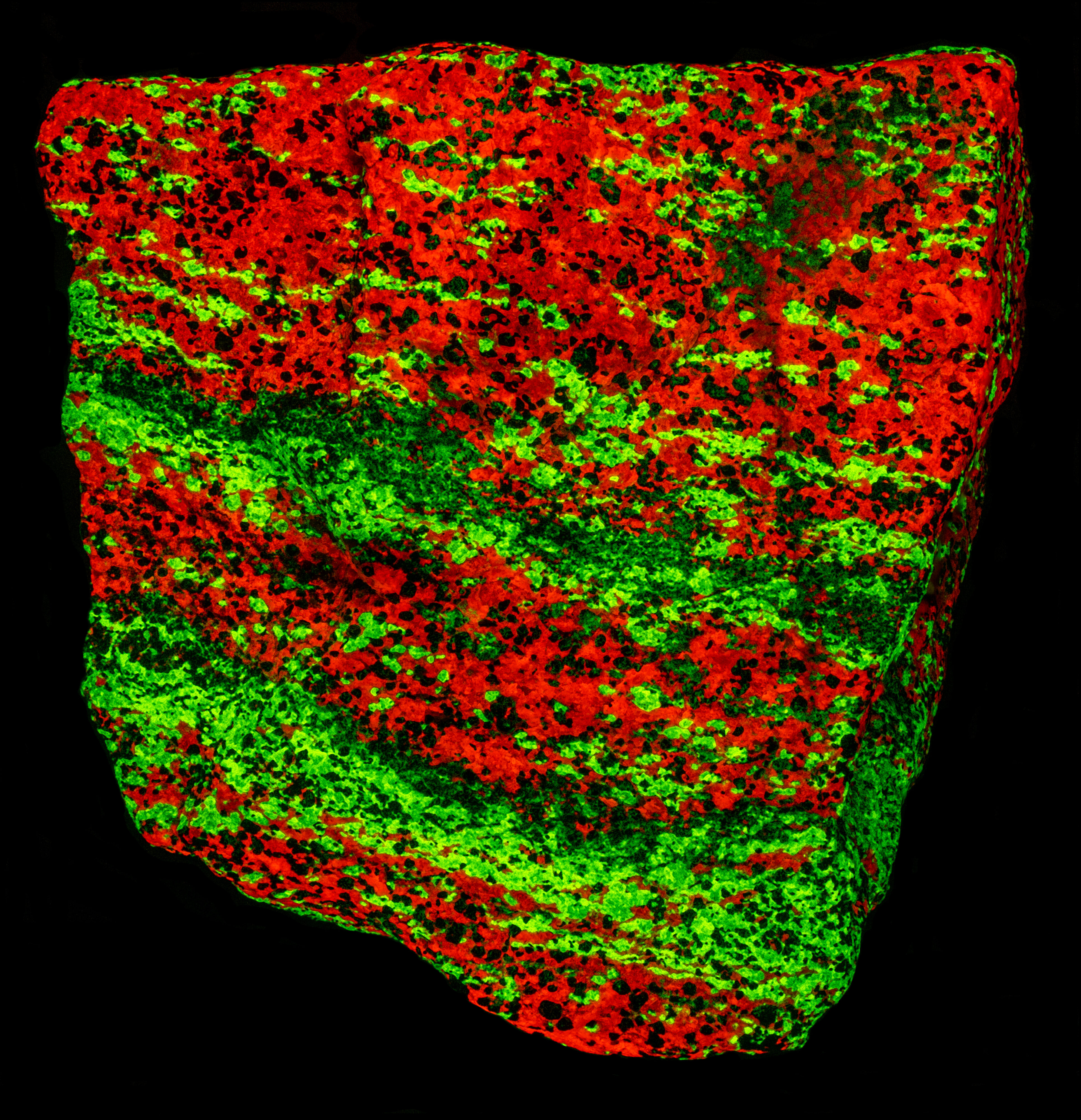
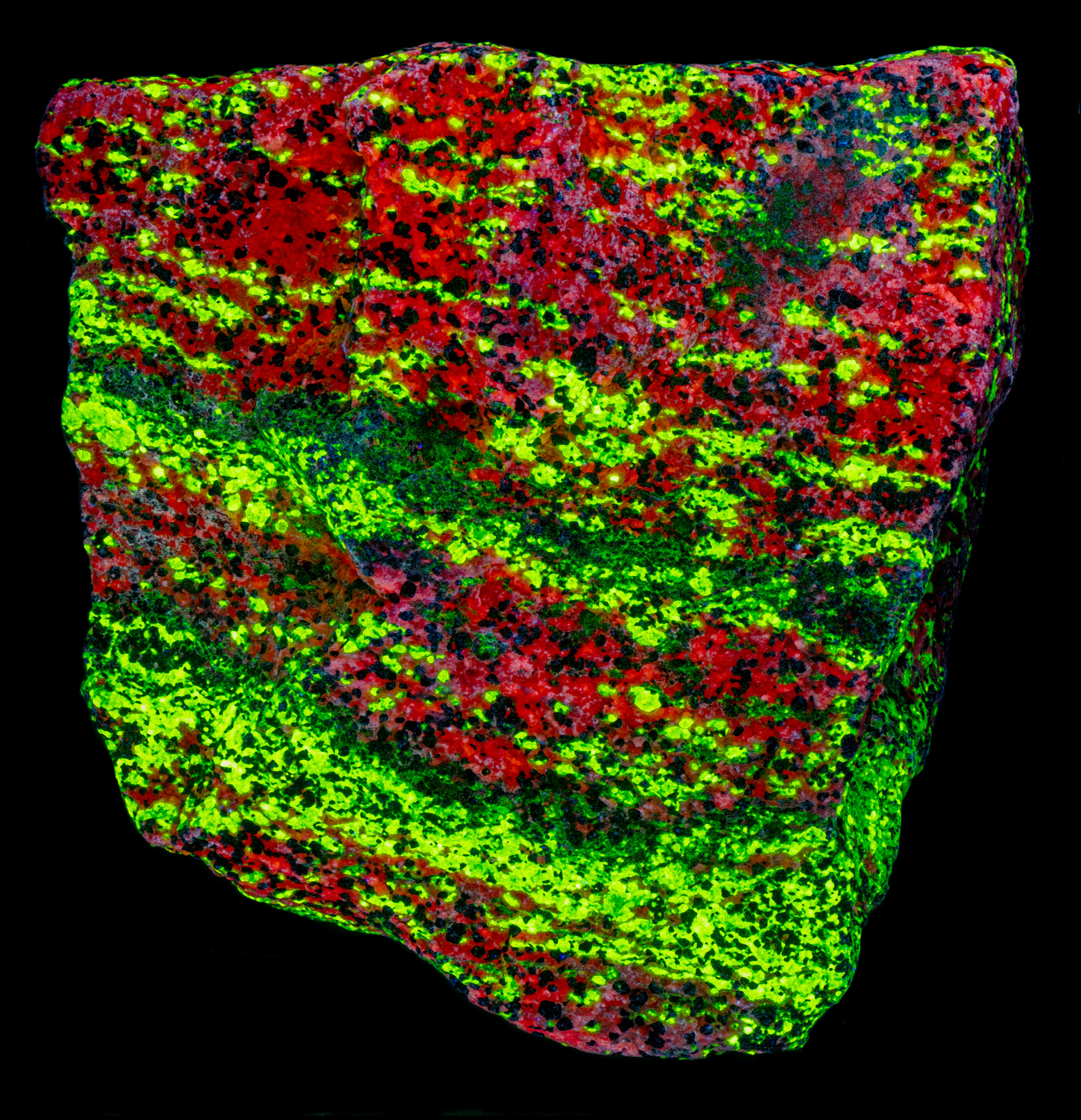
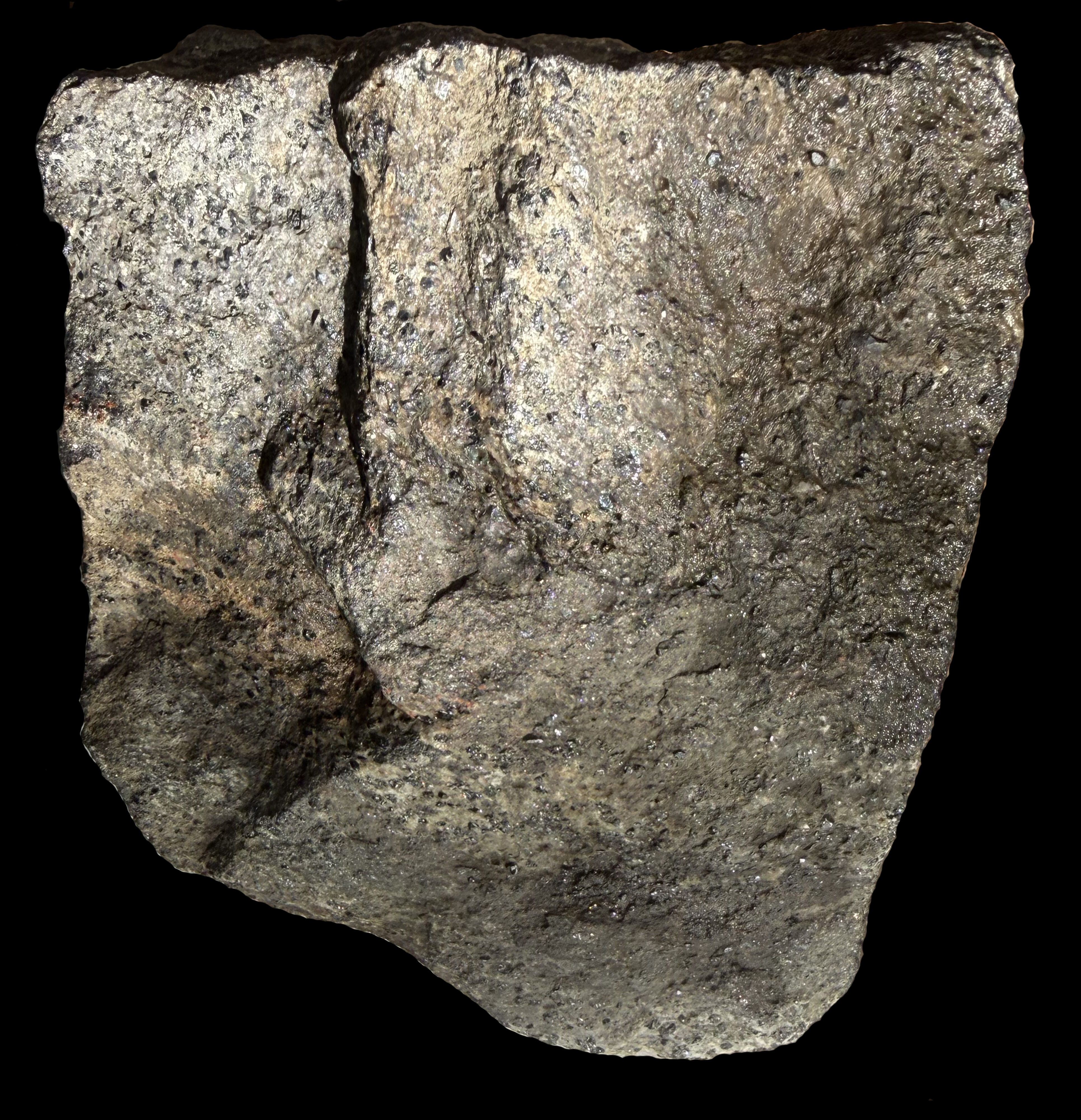
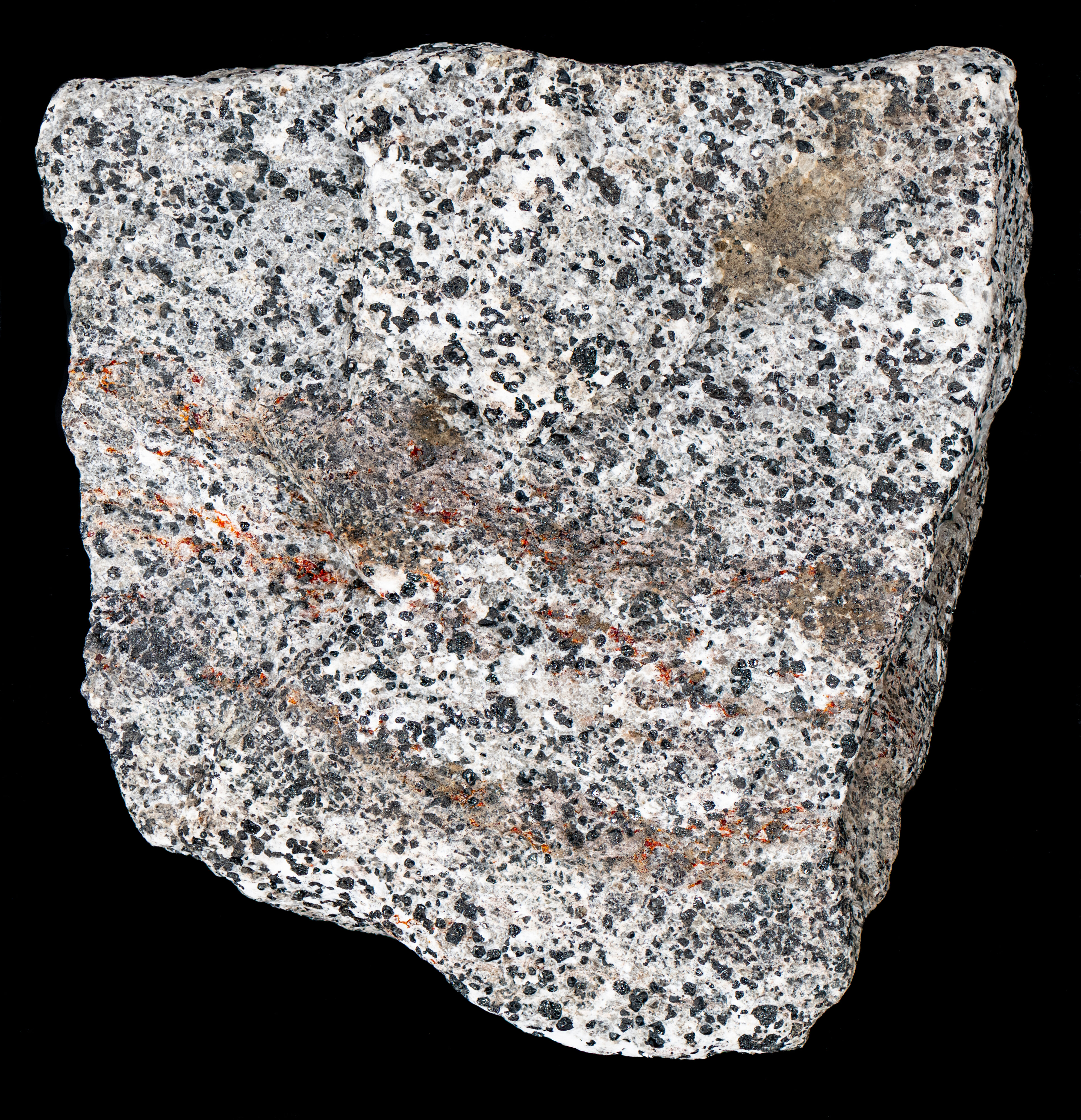
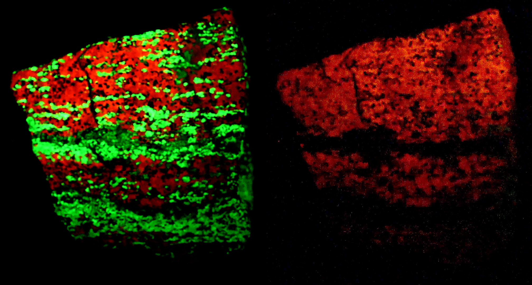
An example of the classic mineral assemblage of green fluorescing willemite (Zn2SiO4), red fluorescing calcite (CaCO3) and non-fluorescent black franklinite (Zn2+Fe3+2O4) and red zincite (ZnO) from the Sterling Mine, Ogdensburg, Sussex County, New Jersey. The surface of this specimen was strongly weathered when I got this specimen. The fluorescence could be seen even with the coating of manganese oxide created by weathering. Unfortunately, I did not take a picture of this fluorescence before I cleaned off the coating. I made a solution of equal parts of 3 percent hydrogen peroxide and white vinegar and placed the rock in it for a couple of hours. The white light pictures show the before and after pictures of the specimen. Banding in the specimen can be seen in the after picture. The banding is more clearly seen in the picture of shortwave fluorescence.
The banding is from the deposition of layers of zinc minerals precipitated from hot brines discharged onto the floor of a shallow sea 1.3 billion years ago. Carbonate muds were also being continuously deposited in this shallow sea. Episodic releases of the hot metalliferous brine into the constant deposition of carbonate muds formed the bands. The original mineralogy that precipitated from the brines is uncertain. The zinc deposits were buried to a depth of 15 to 18 km and heated to temperatures of 700oC to 800oC. These high temperatures and pressures caused high-grade metamorphism of the original mineralogy and left the zinc minerals (willemite, franklinite, and zincite) found in this specimen.
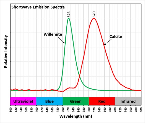
The fluorescence of willemite and calcite is brightest under shortwave illumination. The minerals also fluoresce under longwave and midwave light. The fluorescence in both minerals is activated by manganese. Manganese (Mn2+) replaces zinc in willemite, and manganese replaces calcium in calcite. Shortwave emission spectra show the peak of willemite is at 523 nm and the calcite peak is broader with a maximum at 620 nm. The manganese in the calcite causes red Brief Intense Phosphorescence (BIP) after exposure to all UV wavelengths. There is no afterglow from the willemite.
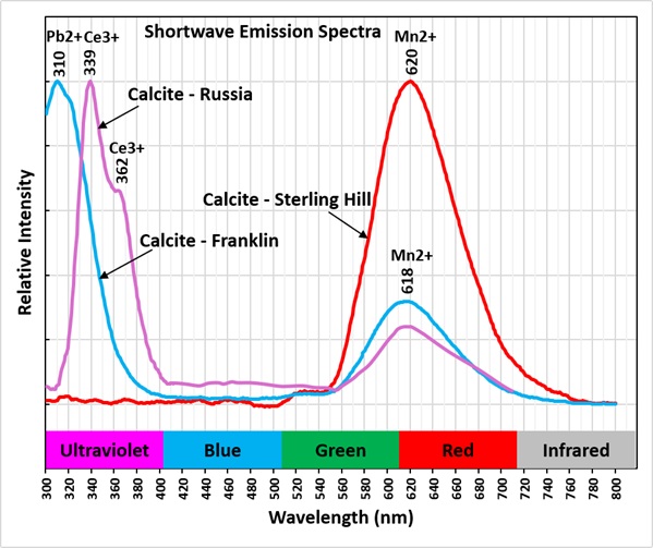
Studies have shown that the red fluorescence of calcite under shortwave light needs a co-activator such as lead (Pb2+) or cerium (Ce3+) along with manganese (Mn2+). These elements replace calcium in the calcite structure. A shortwave emission spectrum of a calcite specimen from Franklin that has peaks for lead and manganese activation is shown in the last spectral plot. Lead produces a very bright and sharp peak with a maximum at 310 nm in the ultraviolet region. The spectral plot also shows a cerium and manganese activated emission spectrum for a calcite from Russia.
This calcite from Sterling Hill has no ultraviolet emission, so there is no obvious co-activator like the other two types of calcite. Possibly the ultraviolet fluorescence is totally absorbed by the manganese ion. More analysis is needed to determine how the red fluorescence is activated in the absence of a co-activator.
Summary of luminescence responses:
Calcite (Mindat) (RRUFF)
- Fluorescence under Longwave (365nm LED) UV light: Red
- Fluorescence under Shortwave (255nm LED) UV light: Red
- Fluorescence under Longwave (365nm LED) UV light: Green
- Fluorescence under Shortwave (255nm LED) UV light: Green