
Scheelite, Calcite and Fluorite - Yaogangxian Mine, Hunan Province, China
Contributed by: Michael Crawford
Date: Jun 25th, 2026
Locality: Yaogangxian Mine, Yaogangxian W-Sn ore field, Yizhang Co., Chenzhou, Hunan, China (See on Mindat)
Size: 4 x 2.5 x 6 cm
Weight: 163 g
Description:
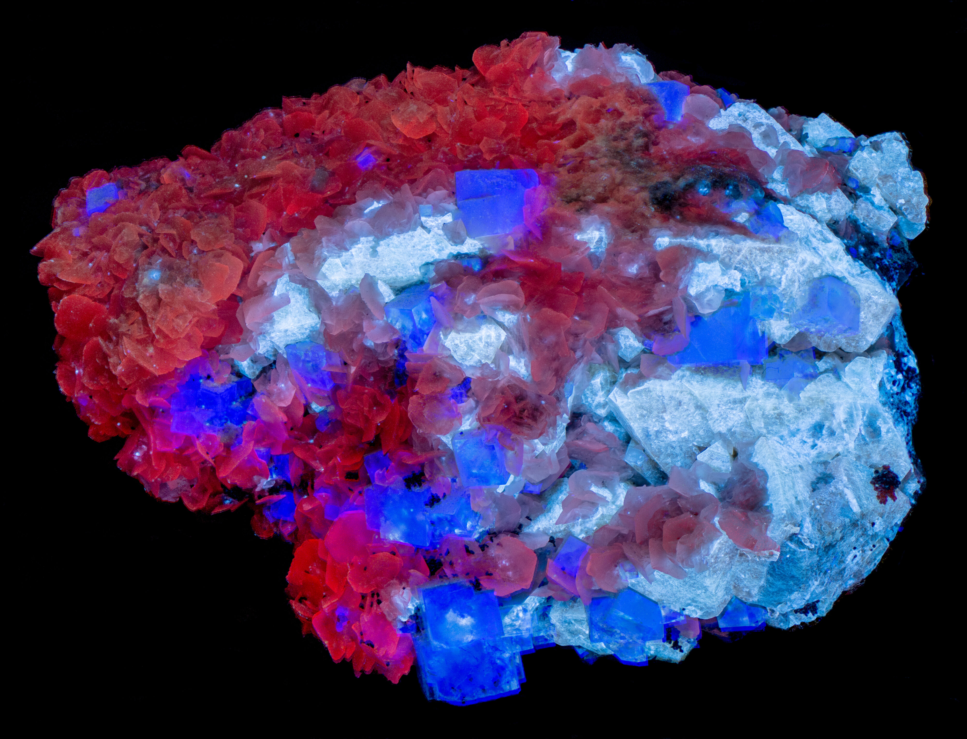
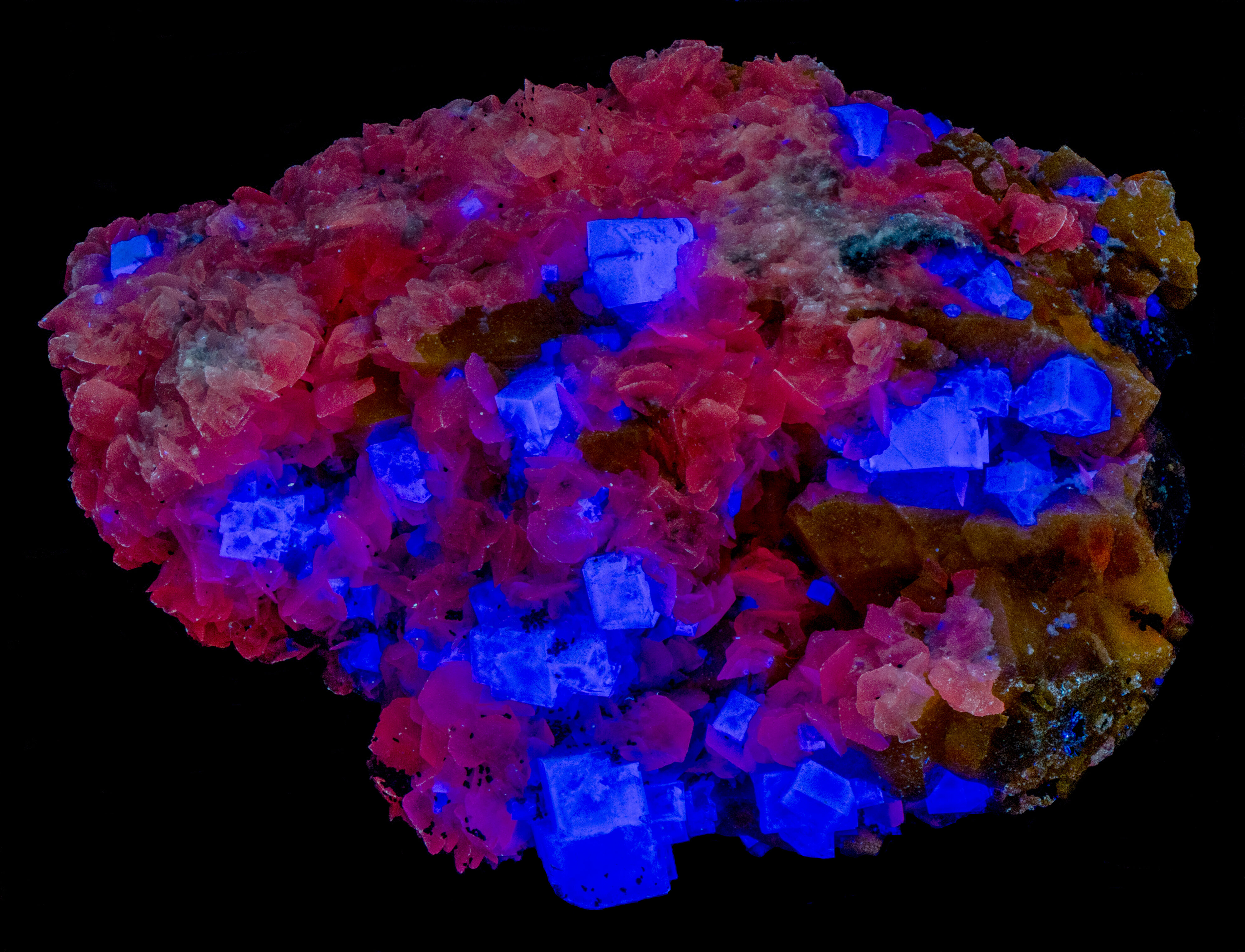
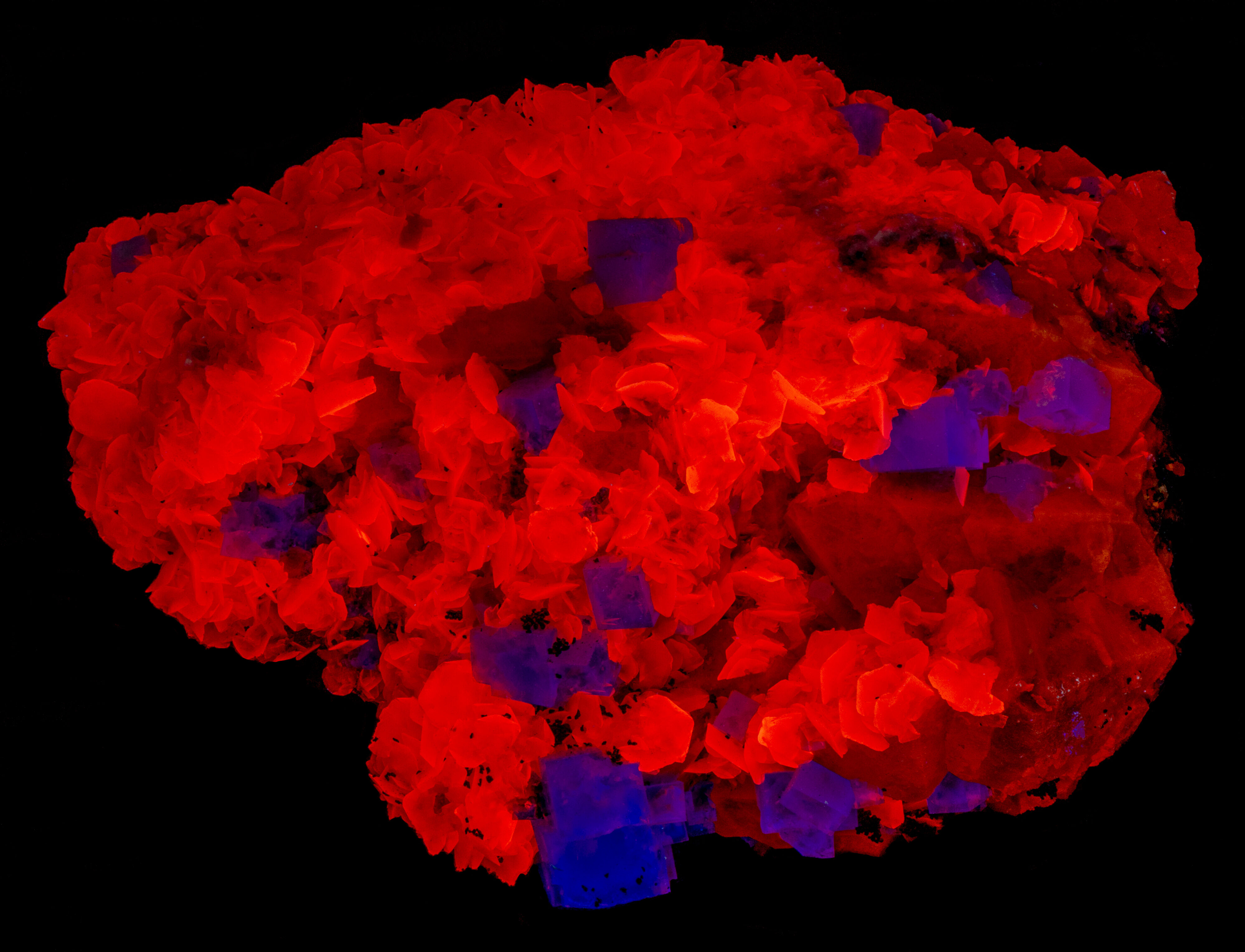
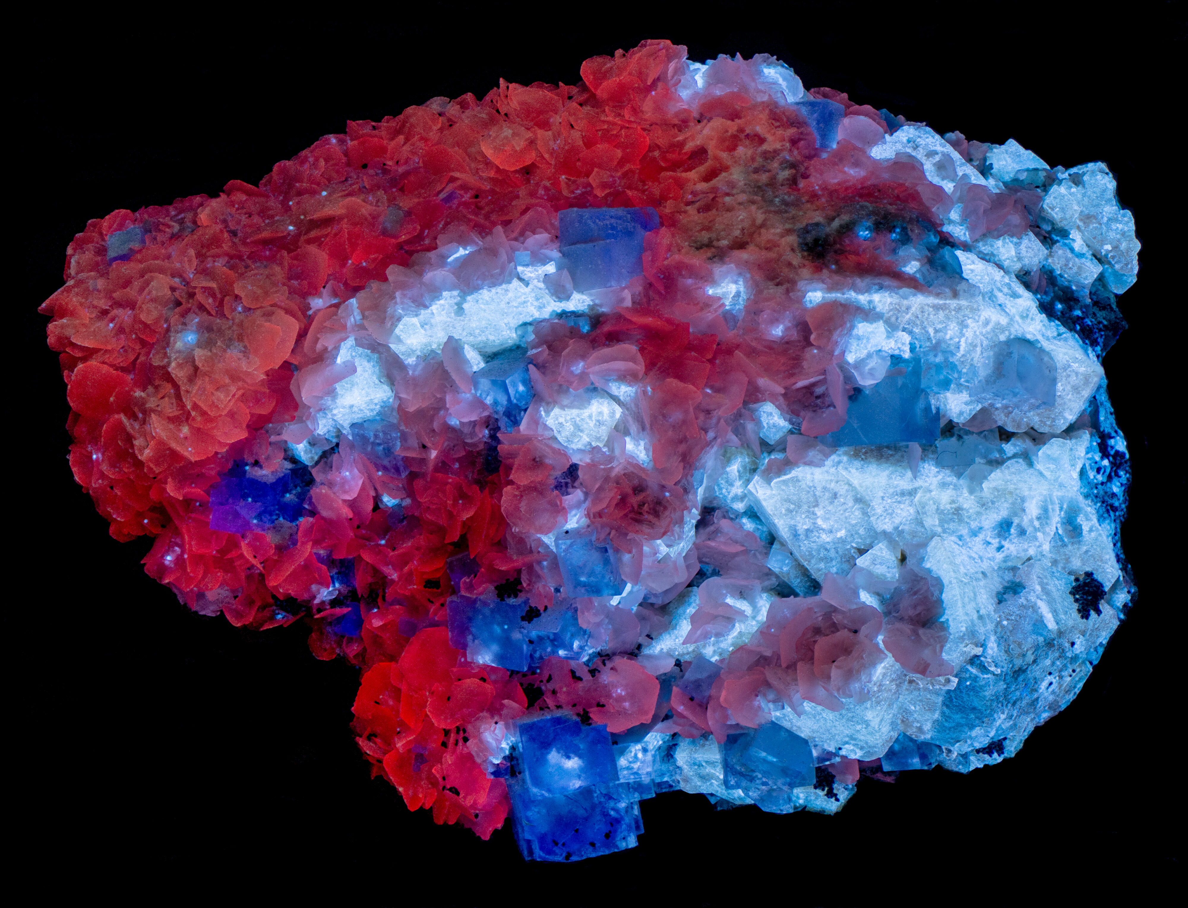
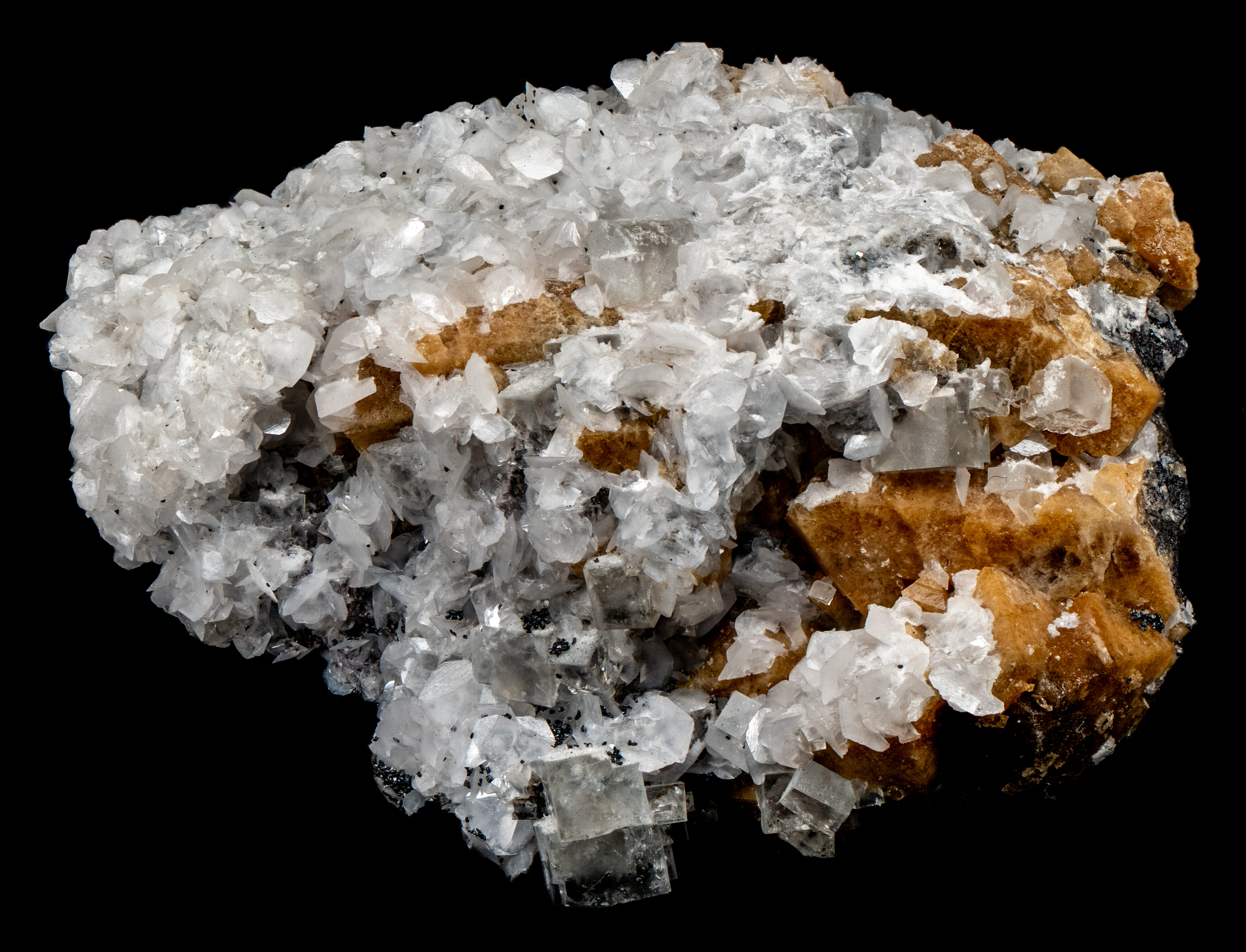
A patriotic red, white and blue specimen for America's 250th> birthday. The specimen was "Made in China". It comes from the Yaogangxian Mine, Hunan Province, China. The "fullwave" image (LW + MW + SW) shows the three fluorescent minerals at their brightest. Blue, fluorescent fluorite (CaF2) is brightest under LW, the red calcite (CaCO3) is brightest under MW, and the scheelite (Ca(WO4)) is brightest under SW. The fluorescent minerals formed on a matrix of black, non-fluorescent wolframite minerals (ferberite (FeWO4) - hubnerite (MnWO4) solid solution).
The fluorescent colors of the fluorite and calcite remain the same under all wavelengths of UV illumination. However, the fluorescent color of the scheelite changes with different wavelengths of UV illumination. The scheelite fluoresces light brown under LW, dark red under MW, and light blue under SW.
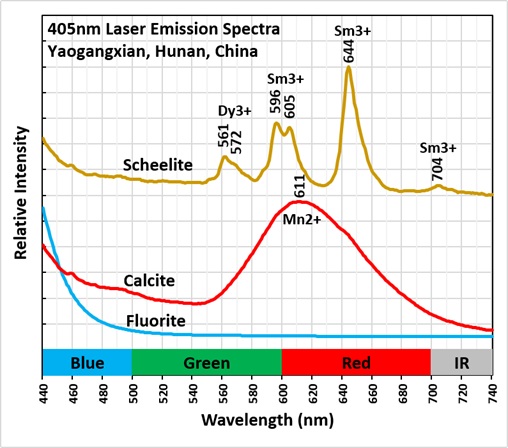
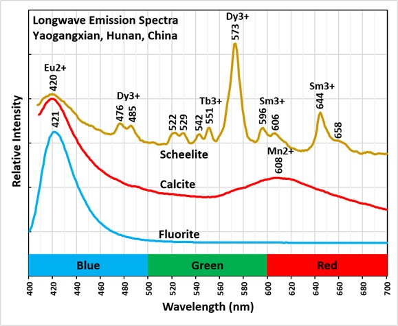
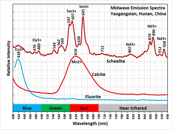
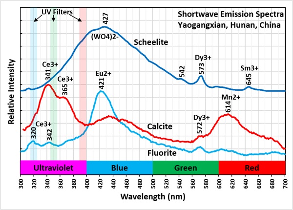
Emission spectra measured with different wavelengths of UV illumination show how the fluorite and calcite remain the same and why scheelite fluorescence changes. Under LW, MW and SW, fluorite has a peak around 420 nm. This peak is activated by europium (Eu2+) replacing calcium in the fluorite structure. Fluorite has slight ultraviolet fluorescence under SW light that is activated by cerium (Ce3+). The peaks at 320 nm and 342 nm are caused by cerium activation.
Red calcite fluorescence is activated by manganese (Mn2+) replacing calcium. This causes a broad peak with a maximum around 608 nm to 616 nm. Under SW, calcite fluoresces in the ultraviolet due to cerium activation. The ultraviolet emission has two sharp peaks at 341 nm and 365 nm caused by the cerium. Cerium is a co-activator for the red fluorescence of calcite under SW light. The ultraviolet fluorescence excites the manganese activated red fluorescence. It is an internal LW light.
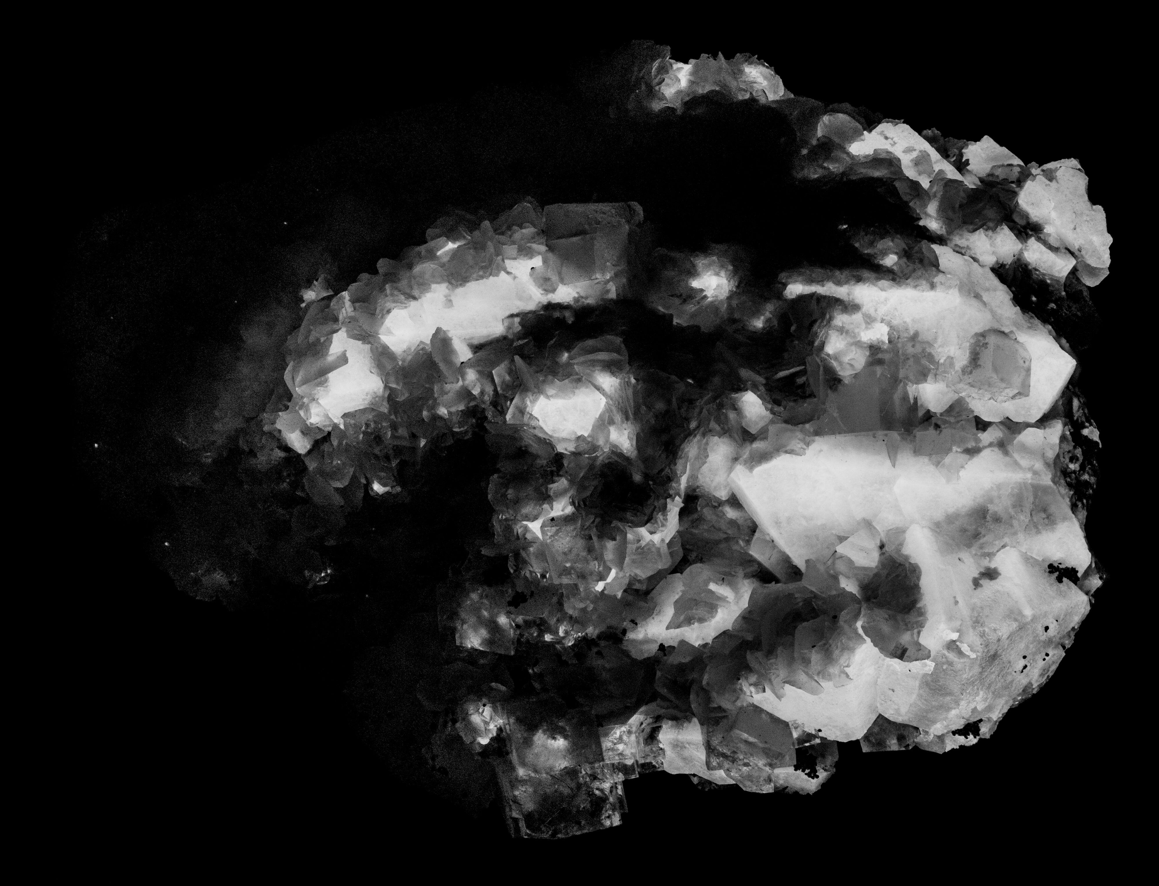
The changing fluorescent colors of scheelite are due to several different elements and ions that are activated by different wavelengths of UV light. The rare earth elements dysprosium (Dy3+) and samarium (Sm3+) are responsible for the color differences under LW and MW light and the tungstate ion ((WO4)2-) causes intrinsic SW fluorescence. The LW light brown fluorescence is dominated by a sharp dysprosium peak at 573 nm and smaller samarium peaks at 596 nm, 606 nm and 644 nm. The red MW fluorescence is dominated by sharp samarium peaks at 597 nm, 607 nm, and 645 nm. There is also strong near infrared fluorescence activated by neodymium (Nd3+) with sharp peaks at 879 nm, 890 nm and 928 nm. This near infrared fluorescence under MW light is clearly shown in the image taken with an 850 nm cutoff filter. The cutoff filter sees any fluorescence with wavelengths longer than 850 nm. Images taken with 850 nm cutoff filter with LW and SW illumination also show the near infrared fluorescence of the scheelite, but the intensity is much less than the MW intensity. The LW and SW near infrared is too dim to be measured with the spectrometer, but bright enough to be imaged by the camera.
The shortwave, blue white fluorescence of scheelite is intrinsic. It is activated by the tungstate ion ((WO4)2-) that is part of scheelite's chemical composition. Axel Ammerman (2010) explained how the tungstate ion activates the blue white scheelite fluorescence:
The crystal structure of scheelite is such that it has tungsten in an eightfold (tetrahedral) coordination within a shell of oxygen atoms. In plain language, tungsten is set in a surrounding of extremely negative oxygen ions. As a result, some of the (negatively charged) electrons that bind tungsten and oxygen are repelled by the (equally negative) crystal field. In other words, they fail to cross the distance off the W-O bond. One could say that the electron is in limbo between two atoms. The positional uncertainty of this electron creates a broad array of energy levels. This broad array of levels translates as a broadband fluorescence. This kind of non-bridging oxygen bonds are denoted as: W-O* in literature (the normal double bond is denoted as W=O.)
The shortwave emission spectrum of the light blue fluorescence is a broad peak with a peak at 427 nm that also extends into the ultraviolet region. The spectrum also contains a couple of small sharp peaks caused by the rare earth elements samarium (Sm3+) and dysprosium (Dy3+) replacing calcium.
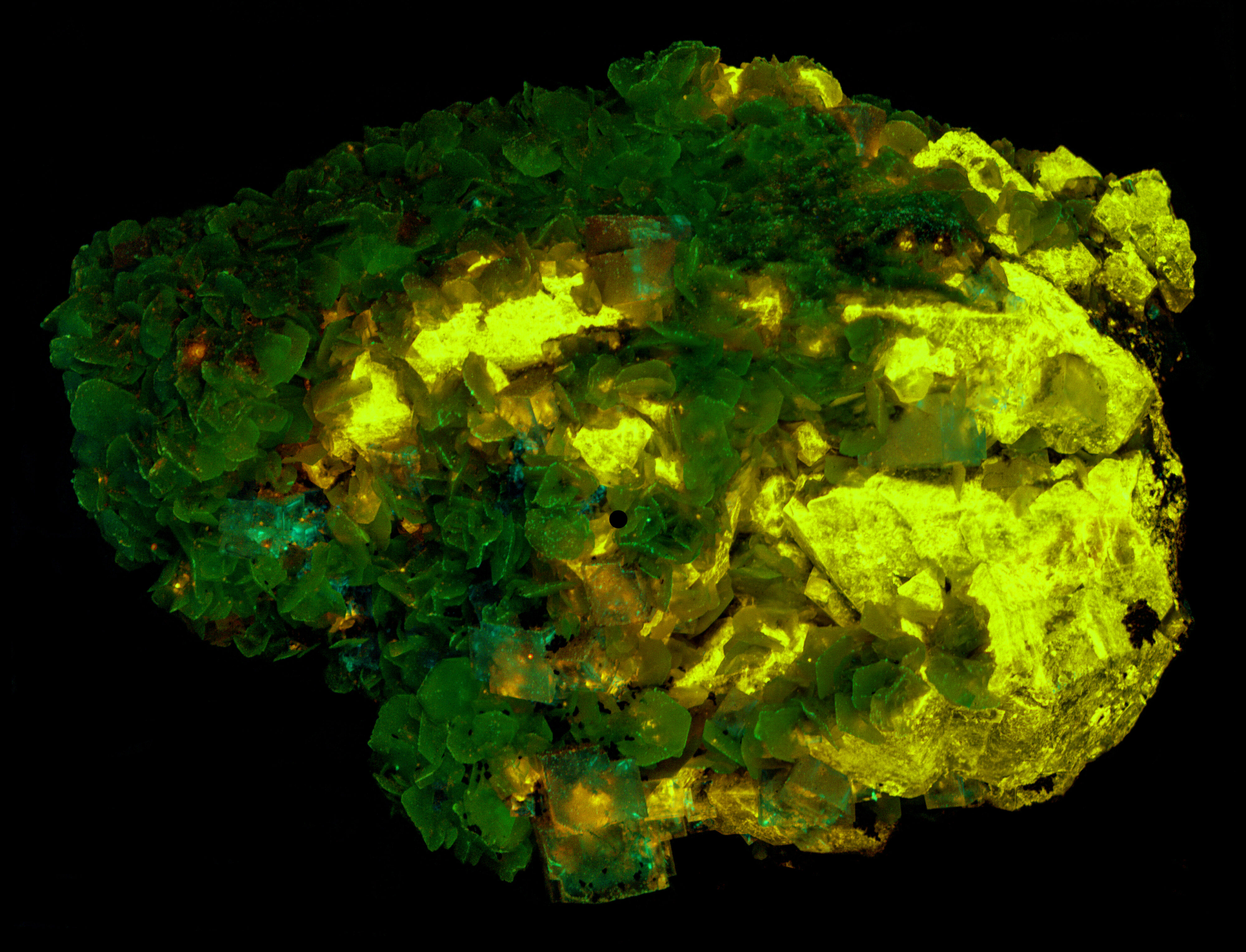
In the false color image of ultraviolet fluorescence, calcite appears green because its ultraviolet fluorescence is brightest in the 350 nm bandpass image (assigned to green). The shoulder of blue scheelite fluorescence extends in the ultraviolet. Scheelite appears yellow in the false color image because it is brightest in the 394 nm image (red) and it moderately bright in the 350 nm image (green). Green plus red creates yellow. Both minerals are non-fluorescent in the 320 nm image, so there is no blue in the image. Ultraviolet fluorescence of fluorite is too dim to be seen in the false color image.
REFERENCE:
Ammerman, A., 2010, Scheelite – not necessarily blue!, Journal of the Fluorescent Mineral Society, V.30, pp. 21-26
Summary of luminescence responses:
Calcite (Mindat) (RRUFF)
- Fluorescence under Longwave (365nm LED) UV light: Red
- Fluorescence under Midwave (305nm LED) UV light: Red
- Fluorescence under Shortwave (255nm LED) UV light: Red
- Fluorescence under Longwave (365nm LED) UV light: Blue
- Fluorescence under Midwave (305nm LED) UV light: Blue
- Fluorescence under Shortwave (255nm LED) UV light: Blue
- Fluorescence under Longwave (365nm LED) UV light: Yellow
- Fluorescence under Midwave (305nm LED) UV light: Red
- Fluorescence under Shortwave (255nm LED) UV light: Blue