Scheelite and Calcite from the Ophir Hill Mine, Tooele County, Utah
Contributed by: Michael Crawford
Date: Oct 1st, 2025
Locality: Ophir Hill Consolidated Mine, Ophir, Ophir Mining District, Tooele County, Utah, USA (See on Mindat)
Size: 7 x 10 cm
Description:
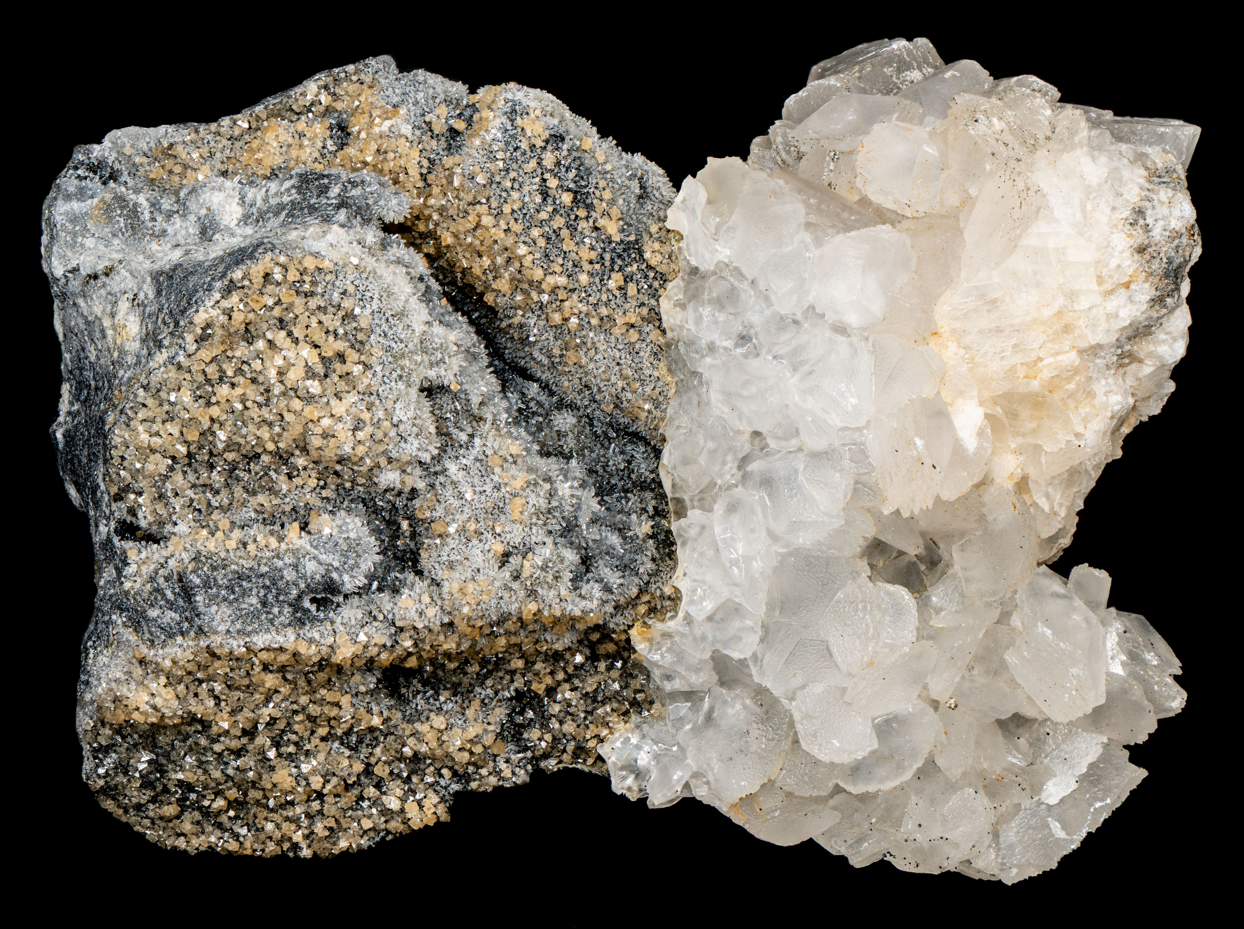
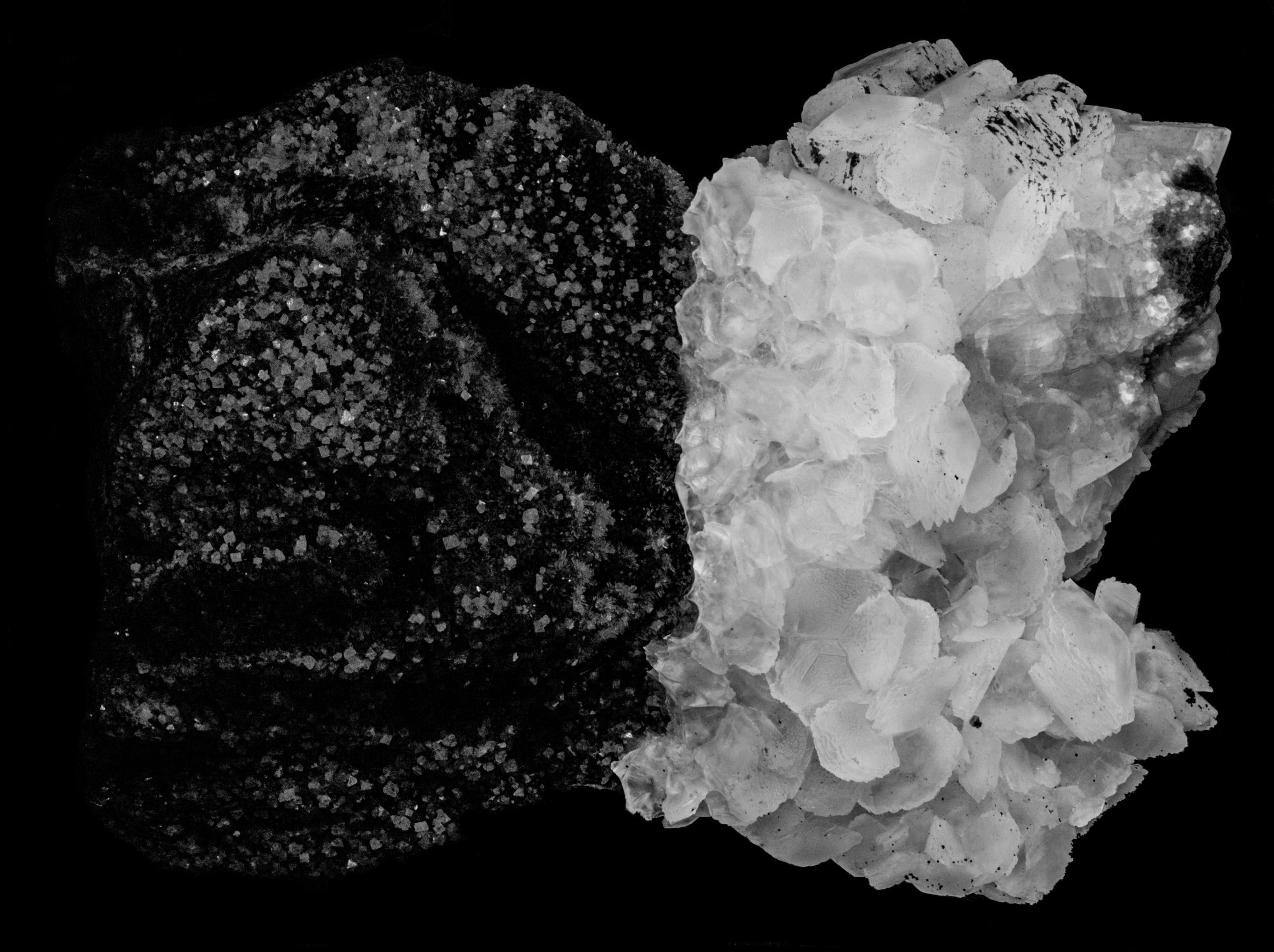
This specimen contains scheelite, quartz, and calcite crystals. The specimen comes from the Ophir Hill Mine, Tooele County, Utah. Mineralization at the Ophir Hill Mine is a replacement polymetallic (Pb-Zn-Ag-Cu-Mn-W) deposit in shaly limestones of Cambrian Ophir Formation, Mississippian Gardison Limestone, and Mississippian lower Great Blue Limestone. The hydrothermal mineralization was deposited along faults and replacement of the faulted limestone.
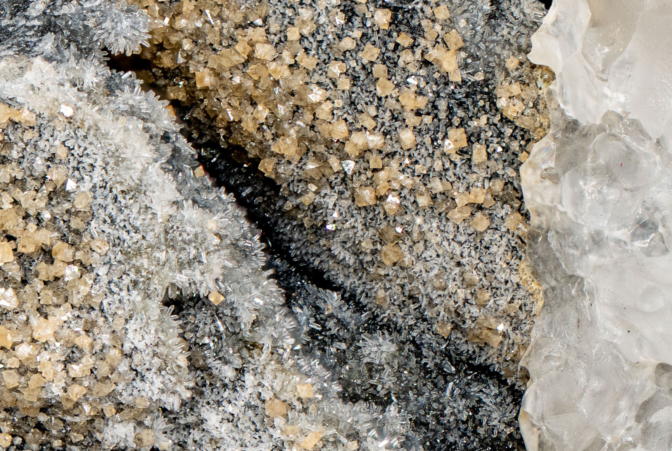
The left half of the specimen contains small yellowish brown scheelite crystals showing tetragonal dipyramid faces and small quartz crystals on a dark massive quartz matrix. The right half of the specimen consists of calcite crystals that cover the scheelite-quartz mineralization. The calcite in the contact area with the scheelite has a smoothed scalloped texture indicating that acid was used to dissolve calcite and expose the scheelite. Scheelite specimens from Ophir Hill Mine are often overgrown with calcite and HCl is used to expose the underlying scheelite.
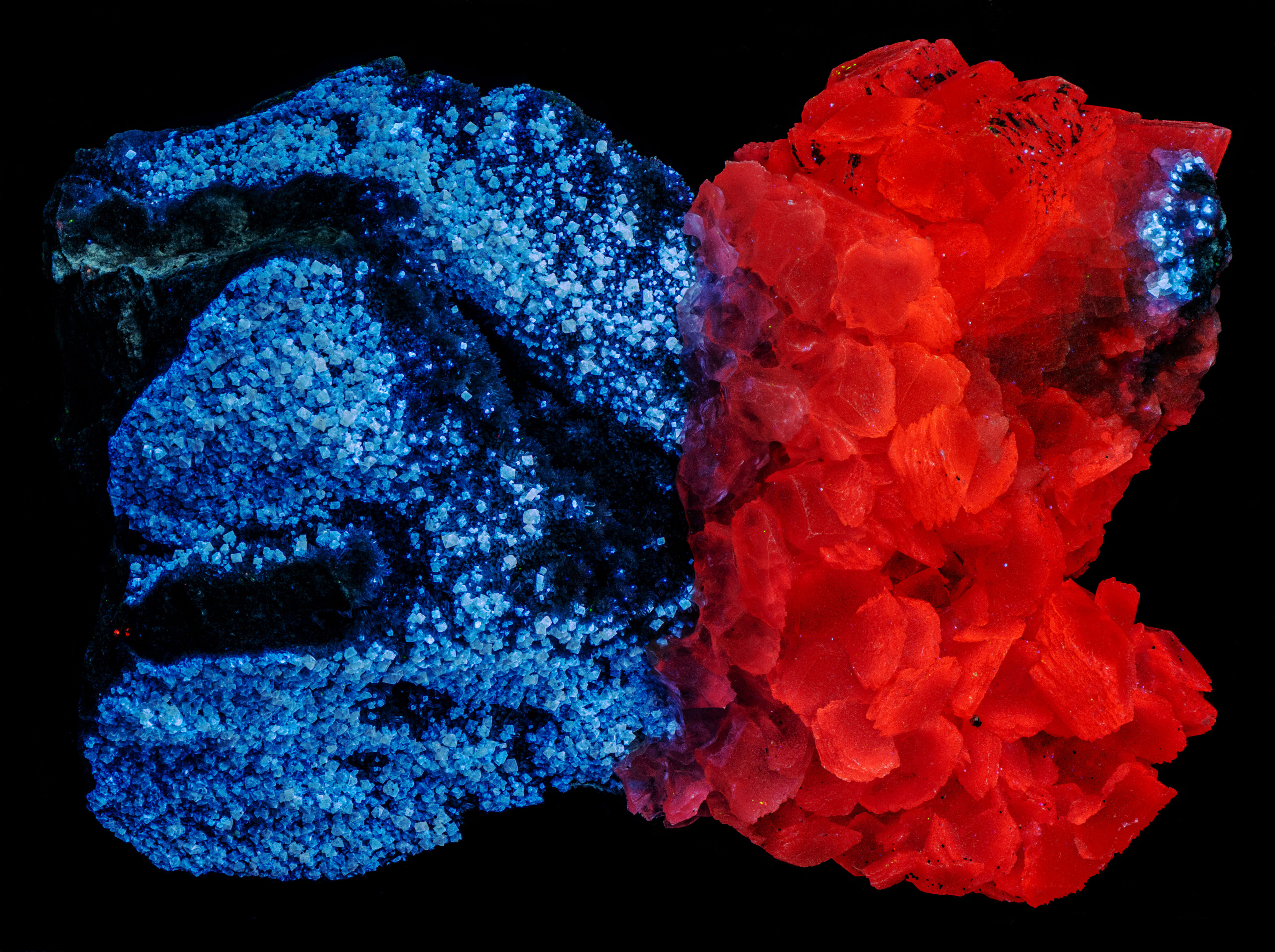
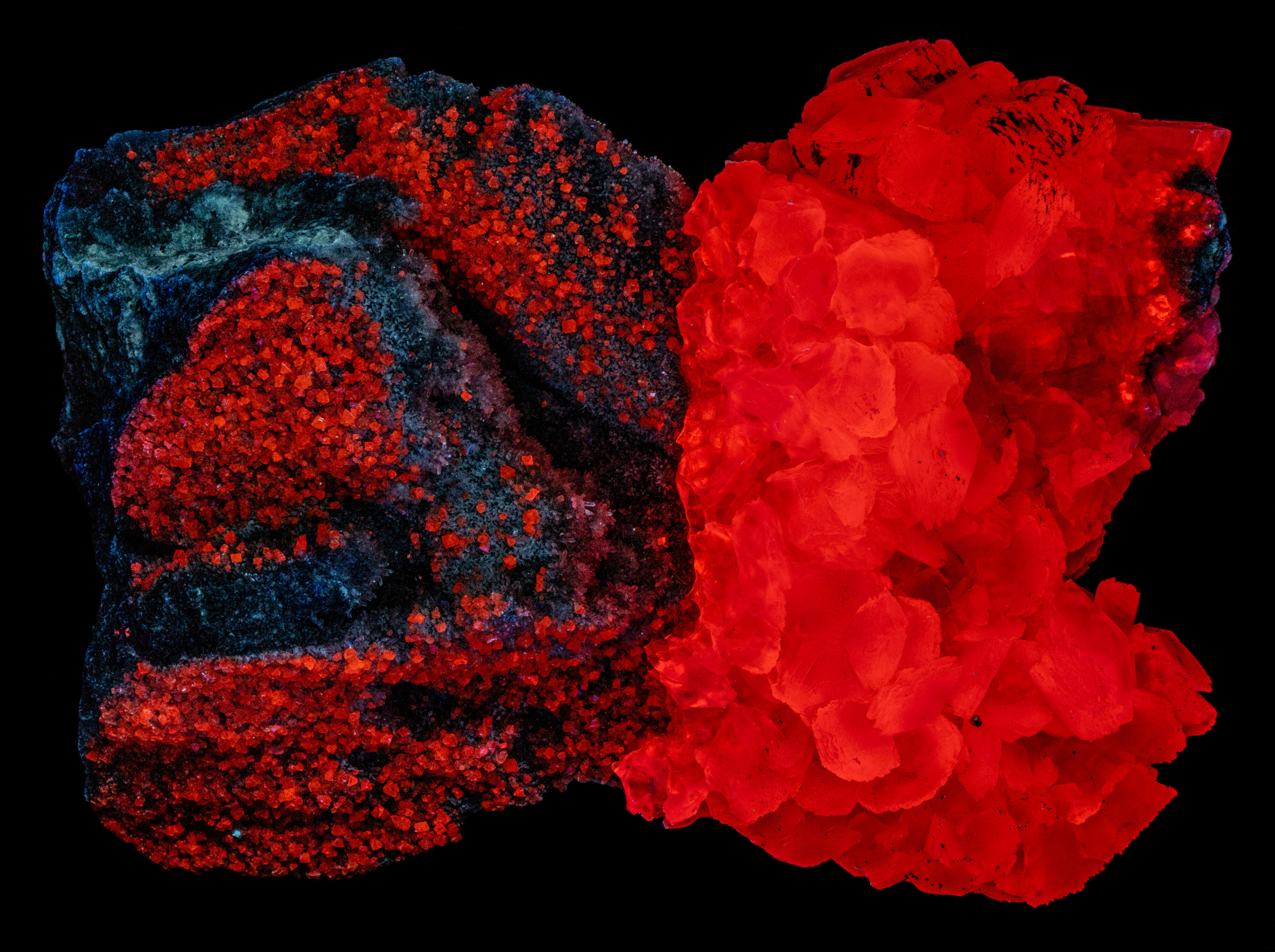
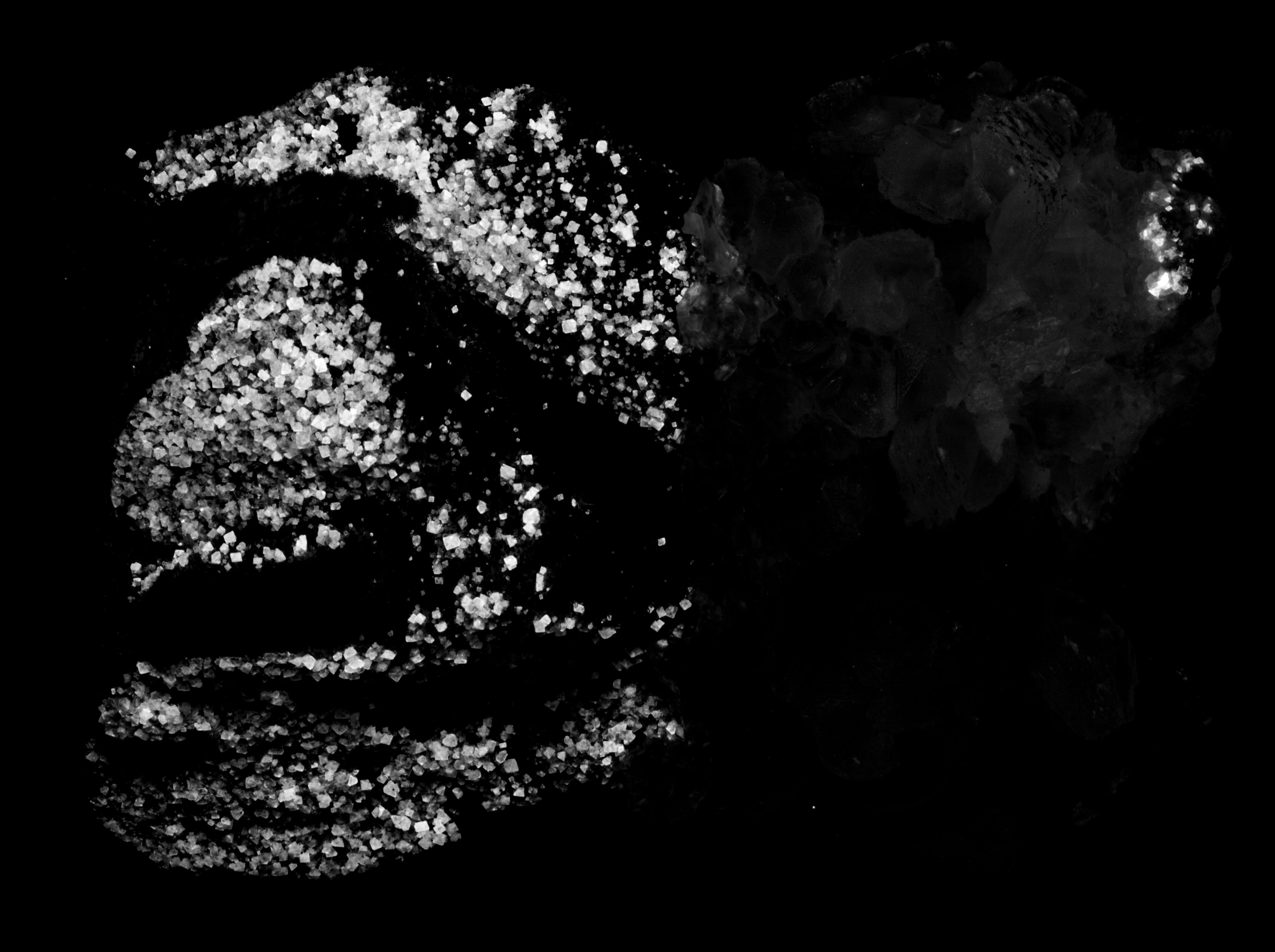
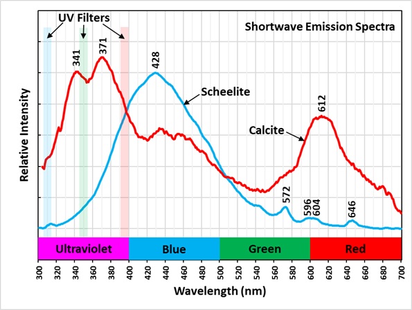
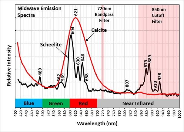
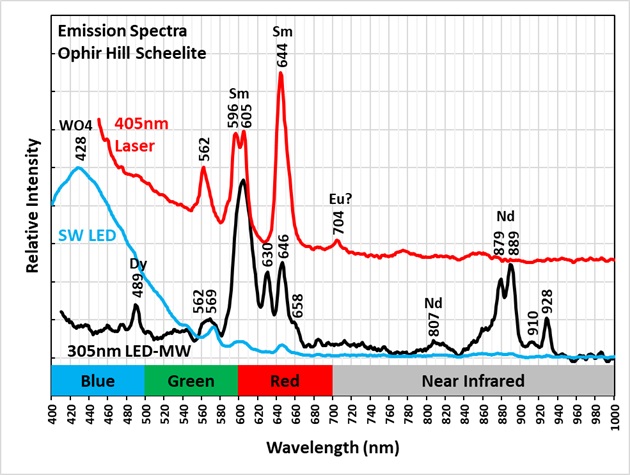
The scheelite has the typical bright bluish white fluorescence under shortwave UV light. This intrinsic fluorescence is activated by the tungstate ion in the scheelite structure. The emission spectrum of this fluorescence peaks at 428 nm and its shoulder extends into the ultraviolet region. The spectrum also contains a couple of small sharp peaks caused by the rare earth elements samarium (Sm) and dysprosium (Dy) replacing calcium. This scheelite’s fluorescence changes to red under midwave light. The tungstate ion no longer activates fluorescence. The midwave emission spectrum contains many sharp peaks in the visible region activated by rare earth elements samarium (Sm) and dysprosium (Dy) that create the red color. The rare earth element, neodymium (Nd), activates near infrared fluorescence. Images of the midwave near infrared fluorescence show the visible fluorescent of calcite extends into the near infrared in the 720 nm bandpass image, but the scheelite is non-fluorescent at this wavelength. The image of the wavelengths longer than 850 nm shows the scheelite fluorescence caused by the neodymium activation. The scheelite is non-fluorescent under longwave light.
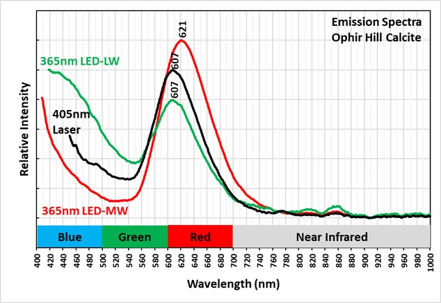
The red calcite fluorescence is activated by manganese replacing calcium. The peak of the emission spectra of the red fluorescence changes with different excitation wavelengths. The peak is at 607 nm under 405 nm laser illumination and 365 nm LED light. The peak is at 621 nm under midwave light (305 nm LED light), and it is at 612 nm under shortwave light (257 nm LED). The shortwave spectrum also shows that the calcite fluoresces in the ultraviolet region. The spectrum has two peaks at 341 nm and 371 nm. This fluorescence is activated by cerium replacing calcium.
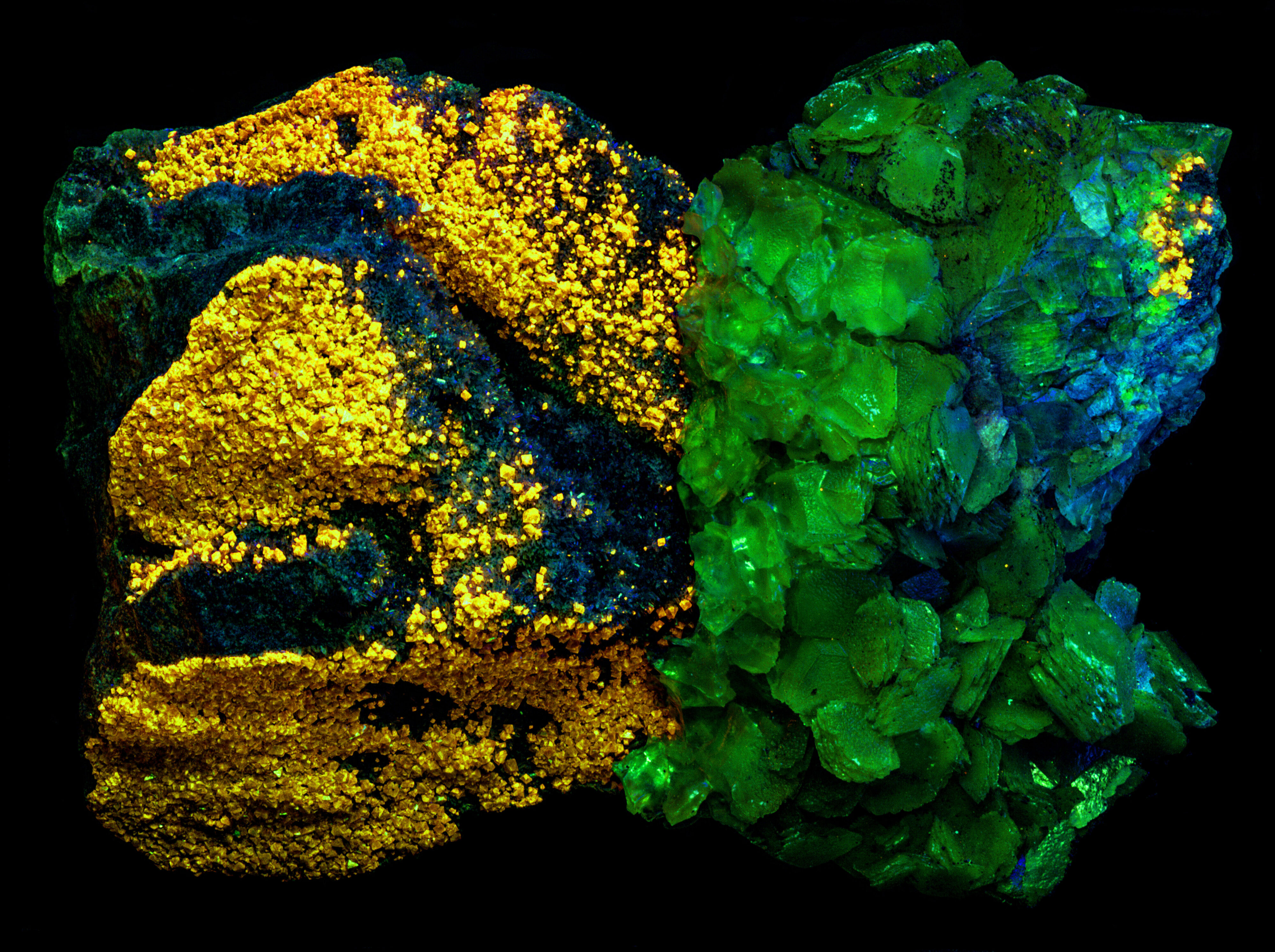
In the false color image of ultraviolet fluorescence, calcite appears green because its ultraviolet fluorescence is brightest in the 350 nm bandpass image (assigned to green). The shoulder of blue scheelite fluorescence extends in the ultraviolet. Scheelite appears yellow in the false color image because it is brightest in the 394 nm image (red) and it moderately bright in the 350 nm image (green). Green plus red creates yellow. Both minerals are non-fluorescent in the 310 nm image, so there is no blue in the image.
Summary of luminescence responses:
Scheelite (Mindat) (RRUFF)
- Fluorescence under Shortwave (255nm LED) UV light: Blue
- Fluorescence under Midwave (305nm LED) UV light: Red
- Fluorescence under Shortwave (255nm LED) UV light: Red
- Fluorescence under Midwave (305nm LED) UV light: Red